49
views
0
recommends
+1 Recommend
0 collections
    0
    shares
      • Record: found
      • Abstract: found
      • Article: not found

      Melody, an ENU mutation in Caspase 3 , alters the catalytic cysteine residue and causes sensorineural hearing loss in mice

      research-article

      Read this article at

      ScienceOpenPublisherPMC
      Bookmark
          There is no author summary for this article yet. Authors can add summaries to their articles on ScienceOpen to make them more accessible to a non-specialist audience.

          Abstract

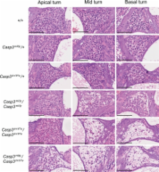
          Progeny from the Harwell N-ethyl- N-nitrosourea (ENU) recessive mutagenesis screen were assessed for auditory defects. A pedigree was identified with multiple progeny lacking response to a clickbox test. Auditory brainstem response (ABR) analysis showed that homozygous mutant mice were profoundly deaf and the line was named melody. We subsequently mapped this mutation to a 6-Mb region on chromosome 8 and identified a point mutation in melody that results in a C163S substitution in the catalytic site of Caspase 3, a cysteine protease involved in apoptosis. Melody fails to complement a null Caspase-3 mutant. Scanning electron microscopy (SEM) has revealed disorganised sensory hair cells and hair cell loss. Histological analysis of melody has shown degeneration of spiral ganglion cells in homozygote mice, with a gradient of severity from apical to basal turns. Melody heterozygotes also show evidence of loss of spiral ganglion neurons, suggesting that the C163S mutation may show dominant negative effects by binding and sequestering proteins at the active site. The melody line provides a new model for studying the role of Caspase 3 in deafness and a number of other pathways and systems.

          Related collections

          Most cited references21

          • Record: found
          • Abstract: found
          • Article: not found

          Caspases: the executioners of apoptosis.

          Apoptosis is a major form of cell death, characterized initially by a series of stereotypic morphological changes. In the nematode Caenorhabditis elegans, the gene ced-3 encodes a protein required for developmental cell death. Since the recognition that CED-3 has sequence identity with the mammalian cysteine protease interleukin-1 beta-converting enzyme (ICE), a family of at least 10 related cysteine proteases has been identified. These proteins are characterized by almost absolute specificity for aspartic acid in the P1 position. All the caspases (ICE-like proteases) contain a conserved QACXG (where X is R, Q or G) pentapeptide active-site motif. Capases are synthesized as inactive proenzymes comprising an N-terminal peptide (prodomain) together with one large and one small subunit. The crystal structures of both caspase-1 and caspase-3 show that the active enzyme is a heterotetramer, containing two small and two large subunits. Activation of caspases during apoptosis results in the cleavage of critical cellular substrates, including poly(ADP-ribose) polymerase and lamins, so precipitating the dramatic morphological changes of apoptosis. Apoptosis induced by CD95 (Fas/APO-1) and tumour necrosis factor activates caspase-8 (MACH/FLICE/Mch5), which contains an N-terminus with FADD (Fas-associating protein with death domain)-like death effector domains, so providing a direct link between cell death receptors and the caspases. The importance of caspase prodomains in the regulation of apoptosis is further highlighted by the recognition of adapter molecules, such as RAIDD [receptor-interacting protein (RIP)-associated ICH-1/CED-3-homologous protein with a death domain]/CRADD (caspase and RIP adapter with death domain), which binds to the prodomain of caspase-2 and recruits it to the signalling complex. Cells undergoing apoptosis following triggering of death receptors execute the death programme by activating a hierarchy of caspases, with caspase-8 and possibly caspase-10 being at or near the apex of this apoptotic cascade.
            Bookmark
            • Record: found
            • Abstract: found
            • Article: not found

            Genome-wide, large-scale production of mutant mice by ENU mutagenesis.

            In the post-genome era, the mouse will have a major role as a model system for functional genome analysis. This requires a large number of mutants similar to the collections available from other model organisms such as Drosophila melanogaster and Caenorhabditis elegans. Here we report on a systematic, genome-wide, mutagenesis screen in mice. As part of the German Human Genome Project, we have undertaken a large-scale ENU-mutagenesis screen for dominant mutations and a limited screen for recessive mutations. In screening over 14,000 mice for a large number of clinically relevant parameters, we recovered 182 mouse mutants for a variety of phenotypes. In addition, 247 variant mouse mutants are currently in genetic confirmation testing and will result in additional new mutant lines. This mutagenesis screen, along with the screen described in the accompanying paper, leads to a significant increase in the number of mouse models available to the scientific community. Our mutant lines are freely accessible to non-commercial users (for information, see http://www.gsf.de/ieg/groups/enu-mouse.html).
              Bookmark
              • Record: found
              • Abstract: found
              • Article: not found

              Calpain and caspase: can you tell the difference?

              Kai Wang (1999)
              Both necrotic and apoptotic neuronal death are observed in various neurological and neurodegenerative disorders. Calpain is activated in various necrotic and apoptotic conditions, while caspase 3 is only activated in neuronal apoptosis. Despite the difference in cleavage-site specificity, an increasing number of cellular proteins are found to be dually susceptible to these cysteine proteases. These include alpha- and beta-fodrin, calmodulin-dependent protein kinases, ADP-ribosyltransferase (ADPRT/PARP) and tau. Intriguingly, calpastatin is susceptible to caspase-mediated fragmentation. Neurotoxic challenges such as hypoxia-hypoglycemia, excitotoxin treatment or metabolic inhibition of cultured neurons result in activation of both proteases. Calpain inhibitors can protect against necrotic neuronal death and, to a lesser extent, apoptotic death. Caspase inhibitors strongly suppress apoptotic neuronal death. Thus, both protease families might contribute to structural derangement and functional loss in neurons under degenerative conditions.
                Bookmark

                Author and article information

                Contributors
                s.brown@har.mrc.ac.uk
                Journal
                Mamm Genome
                Mammalian Genome
                Springer-Verlag (New York )
                0938-8990
                1432-1777
                30 November 2010
                30 November 2010
                December 2010
                : 21
                : 11-12
                : 565-576
                Affiliations
                MRC Mammalian Genetics Unit, MRC Harwell, Harwell Science and Innovation Campus, Oxfordshire, OX11 0RD UK
                Article
                9306
                10.1007/s00335-010-9306-2
                3002157
                21116635
                356b86e3-5465-4138-a07e-a4c9b4eefc6a
                © The Author(s) 2010
                History
                : 22 April 2010
                : 8 November 2010
                Categories
                Article
                Custom metadata
                © Springer Science+Business Media, LLC 2010

                Genetics
                Genetics

                Comments

                Comment on this article