13
views
0
recommends
+1 Recommend
0 collections
    0
    shares
      • Record: found
      • Abstract: found
      • Article: found
      Is Open Access

      Porcine Bladder Urothelial, Myofibroblast, and Detrusor Muscle Cells: Characterization and ATP Release

      research-article

      Read this article at

      Bookmark
          There is no author summary for this article yet. Authors can add summaries to their articles on ScienceOpen to make them more accessible to a non-specialist audience.

          Abstract

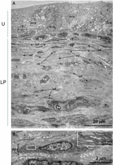
          ATP is released from the bladder mucosa in response to stretch, but the cell types responsible are unclear. Our aim was to isolate and characterize individual populations of urothelial, myofibroblast, and detrusor muscle cells in culture, and to examine agonist-stimulated ATP release. Using female pig bladders, urothelial cells were isolated from bladder mucosa following trypsin-digestion of the luminal surface. The underlying myofibroblast layer was dissected, minced, digested, and cultured until confluent (10–14 days). A similar protocol was used for muscle cells. Cultures were used for immunocytochemical staining and/or ATP release investigations. In urothelial cultures, immunoreactivity was present for the cytokeratin marker AE1/AE3 but not the contractile protein α-smooth muscle actin (α-SMA) or the cytoskeletal filament vimentin. Neither myofibroblast nor muscle cell cultures stained for AE1/AE3. Myofibroblast cultures partially stained for α-SMA, whereas muscle cultures were 100% stained. Both myofibroblast and muscle stained for vimentin, however, they were morphologically distinct. Ultrastructural studies verified that the suburothelial layer of pig bladder contained abundant myofibroblasts, characterized by high densities of rough endoplasmic reticulum. Baseline ATP release was higher in urothelial and myofibroblast cultures, compared with muscle. ATP release was significantly stimulated by stretch in all three cell populations. Only urothelial cells released ATP in response to acid, and only muscle cells were stimulated by capsaicin. Tachykinins had no effect on ATP release. In conclusion, we have established a method for culture of three cell populations from porcine bladder, a well-known human bladder model, and shown that these are distinct morphologically, immunologically, and pharmacologically.

          Related collections

          Most cited references42

          • Record: found
          • Abstract: found
          • Article: not found

          Altered urinary bladder function in mice lacking the vanilloid receptor TRPV1.

          In the urinary bladder, the capsaicin-gated ion channel TRPV1 is expressed both within afferent nerve terminals and within the epithelial cells that line the bladder lumen. To determine the significance of this expression pattern, we analyzed bladder function in mice lacking TRPV1. Compared with wild-type littermates, trpv1(-/-) mice had a higher frequency of low-amplitude, non-voiding bladder contractions. This alteration was accompanied by reductions in both spinal cord signaling and reflex voiding during bladder filling (under anesthesia). In vitro, stretch-evoked ATP release and membrane capacitance changes were diminished in bladders excised from trpv1(-/-) mice, as was hypoosmolality-evoked ATP release from cultured trpv1(-/-) urothelial cells. These findings indicate that TRPV1 participates in normal bladder function and is essential for normal mechanically evoked purinergic signaling by the urothelium.
            Bookmark
            • Record: found
            • Abstract: not found
            • Article: not found

            Vanilloid receptor expression suggests a sensory role for urinary bladder epithelial cells.

              Bookmark
              • Record: found
              • Abstract: found
              • Article: not found

              Morphology and localization of interstitial cells in the guinea pig bladder: structural relationships with smooth muscle and neurons.

              In the current study we examined the location of interstitial cell of Cajal (ICC)-like cells in the guinea pig bladder wall and studied their structural interactions with nerves and smooth muscle cells. Whole mount samples and cryosections of bladder tissue were labeled with primary and fluorescent secondary antibodies, and imaged using confocal and multiphoton microscopy. Kit positive ICC-like cells were located below the urothelium, in the lamina propria region and throughout the detrusor. In the suburothelium they had a stellate morphology and appeared to network. They made connections with nerves, as shown by double labeling experiments with anti-kit and anti-protein gene product 9.5. A network of vimentin positive cells was also found, of which many but not all were kit positive. In the detrusor kit positive cells were most often seen at the edge of smooth muscle bundles. They were elongated with lateral branches, running in parallel with the bundles and closely associated with intramural nerves. Another population of kit positive cells was seen in the detrusor between muscle bundles. These cells had a more stellate-like morphology and made connections with each other. Kit positive cells were seen tracking nerve bundles and close to intramural ganglia. Vimentin positive cells were present in the detrusor, of which some were also kit positive. There are several populations of ICC-like cells throughout the guinea pig bladder wall. They differ in morphology and orientation but all make connections with intramural nerves and in the detrusor they are closely associated with smooth muscle cells.
                Bookmark

                Author and article information

                Journal
                Front Pharmacol
                Front. Pharmacol.
                Frontiers in Pharmacology
                Frontiers Research Foundation
                1663-9812
                07 June 2011
                2011
                : 2
                : 27
                Affiliations
                [1] 1simpleDetrusor Muscle Laboratory, St George Hospital, University of New South Wales Kogarah, NSW, Australia
                [2] 2simpleGraduate School of Medicine, University of Wollongong Wollongong, NSW, Australia
                [3] 3simpleDepartment of Pharmacology, School of Medical Sciences, University of New South Wales Sydney, NSW, Australia
                Author notes

                Edited by: Bimal Malhotra, Pfizer, USA

                Reviewed by: Peter Ochodnicky, Academic Medical Center Amsterdam, Netherlands; Robert S. Moreland, Drexel University College of Medicine, USA; Robert James Theobald, A. T. Still University of Health Sciences-Kirksville College of Osteopathic Medicine, USA

                *Correspondence: Kylie J. Mansfield, Graduate School of Medicine, University of Wollongong, Northfields Avenue, Wollongong, NSW 2522, Australia. e-mail: kylie_mansfield@ 123456uow.edu.au

                This article was submitted to Frontiers in Cardiovascular and Smooth Muscle Pharmacology, a specialty of Frontiers in Pharmacology.

                Article
                10.3389/fphar.2011.00027
                3113165
                21713125
                373d12cb-3169-49ac-a3ac-93b8ed3f96ce
                Copyright © 2011 Cheng, Mansfield, Sandow, Sadananda, Burcher and Moore.

                This is an open-access article subject to a non-exclusive license between the authors and Frontiers Media SA, which permits use, distribution and reproduction in other forums, provided the original authors and source are credited and other Frontiers conditions are complied with.

                History
                : 13 March 2011
                : 23 May 2011
                Page count
                Figures: 5, Tables: 1, Equations: 0, References: 54, Pages: 9, Words: 6925
                Categories
                Pharmacology
                Original Research

                Pharmacology & Pharmaceutical medicine
                porcine,myofibroblast,detrusor,ultrastructure,urothelium,bladder,atp

                Comments

                Comment on this article