178
views
0
recommends
+1 Recommend
0 collections
    0
    shares
      • Record: found
      • Abstract: found
      • Article: found
      Is Open Access

      Vildagliptin preserves the mass and function of pancreatic β cells via the developmental regulation and suppression of oxidative and endoplasmic reticulum stress in a mouse model of diabetes

      research-article

      Read this article at

      Bookmark
          There is no author summary for this article yet. Authors can add summaries to their articles on ScienceOpen to make them more accessible to a non-specialist audience.

          Abstract

          Aim

          We investigated the molecular mechanisms by which vildagliptin preserved pancreatic β cell mass and function.

          Methods

          Morphological, biochemical and gene expression profiles of the pancreatic islets were investigated in male KK-A y -TaJcl(KK-A y ) and C57BL/6JJcl (B6) mice aged 8 weeks which received either vildagliptin or a vehicle for 4 weeks.

          Results

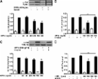
          Body weight, food intake, fasting blood glucose, plasma insulin and active glucagon-like peptide-1 were unchanged with vildagliptin treatment in both mice. In KK-A y mice treated with vildagliptin, increased plasma triglyceride (TG) level and islet TG content were decreased, insulin sensitivity significantly improved, and the glucose tolerance ameliorated with increases in plasma insulin levels. Furthermore, vildagliptin increased glucose-stimulated insulin secretion, islet insulin content and pancreatic β cell mass in both strains. By vildagliptin, the expression of genes involved in cell differentiation/proliferation was upregulated in both strains, those related to apoptosis, endoplasmic reticulum stress and lipid synthesis was decreased and those related to anti-apoptosis and anti-oxidative stress was upregulated, in KK-A y mice. The morphological results were consistent with the gene expression profiles.

          Conclusion

          Vildagliptin increases β cell mass by not only directly affecting cell kinetics but also by indirectly reducing cell apoptosis, oxidative stress and endoplasmic reticulum stress in diabetic mice.

          Related collections

          Most cited references32

          • Record: found
          • Abstract: found
          • Article: not found

          Preserved incretin activity of glucagon-like peptide 1 [7-36 amide] but not of synthetic human gastric inhibitory polypeptide in patients with type-2 diabetes mellitus.

          In type-2 diabetes, the overall incretin effect is reduced. The present investigation was designed to compare insulinotropic actions of exogenous incretin hormones (gastric inhibitory peptide [GIP] and glucagon-like peptide 1 [GLP-1] [7-36 amide]) in nine type-2 diabetic patients (fasting plasma glucose 7.8 mmol/liter; hemoglobin A1c 6.3 +/- 0.6%) and in nine age- and weight-matched normal subjects. Synthetic human GIP (0.8 and 2.4 pmol/kg.min over 1 h each), GLP-1 [7-36 amide] (0.4 and 1.2 pmol/kg.min over 1 h each), and placebo were administered under hyperglycemic clamp conditions (8.75 mmol/liter) in separate experiments. Plasma GIP and GLP-1 [7-36 amide] concentrations (radioimmunoassay) were comparable to those after oral glucose with the low, and clearly supraphysiological with the high infusion rates. Both GIP and GLP-1 [7-36 amide] dose-dependently augmented insulin secretion (insulin, C-peptide) in both groups (P < 0.05). With GIP, the maximum effect in type-2 diabetic patients was significantly lower (by 54%; P < 0.05) than in normal subjects. With GLP-1 [7-36 amide] type-2 diabetic patients reached 71% of the increments in C-peptide of normal subjects (difference not significant). Glucagon was lowered during hyperglycemic clamps in normal subjects, but not in type-2 diabetic patients, and further by GLP-1 [7-36 amide] in both groups (P < 0.05), but not by GIP. In conclusion, in mild type-2 diabetes, GLP-1 [7-36 amide], in contrast to GIP, retains much of its insulinotropic activity. It also lowers glucagon concentrations.
            Bookmark
            • Record: found
            • Abstract: found
            • Article: not found

            Dipeptidyl-peptidase IV from bench to bedside: an update on structural properties, functions, and clinical aspects of the enzyme DPP IV.

            Dipeptidyl-peptidase IV/CD26 (DPP IV) is a cell-surface protease belonging to the prolyloligopeptidase family. It selectively removes the N-terminal dipeptide from peptides with proline or alanine in the second position. Apart from its catalytic activity, it interacts with several proteins, for instance, adenosine deaminase, the HIV gp120 protein, fibronectin, collagen, the chemokine receptor CXCR4, and the tyrosine phosphatase CD45. DPP IV is expressed on a specific set of T lymphocytes, where it is up-regulated after activation. It is also expressed in a variety of tissues, primarily on endothelial and epithelial cells. A soluble form is present in plasma and other body fluids. DPP IV has been proposed as a diagnostic or prognostic marker for various tumors, hematological malignancies, immunological, inflammatory, psychoneuroendocrine disorders, and viral infections. DPP IV truncates many bioactive peptides of medical importance. It plays a role in glucose homeostasis through proteolytic inactivation of the incretins. DPP IV inhibitors improve glucose tolerance and pancreatic islet cell function in animal models of type 2 diabetes and in diabetic patients. The role of DPP IV/ CD26 within the immune system is a combination of its exopeptidase activity and its interactions with different molecules. This enables DPP IV/CD26 to serve as a co-stimulatory molecule to influence T cell activity and to modulate chemotaxis. DPP IV is also implicated in HIV-1 entry, malignant transformation, and tumor invasion.
              Bookmark
              • Record: found
              • Abstract: found
              • Article: found
              Is Open Access

              Dipeptidyl Peptidase 4 Is a Novel Adipokine Potentially Linking Obesity to the Metabolic Syndrome

              OBJECTIVE Comprehensive proteomic profiling of the human adipocyte secretome identified dipeptidyl peptidase 4 (DPP4) as a novel adipokine. This study assessed the functional implications of the adipokine DPP4 and its association to the metabolic syndrome. RESEARCH DESIGN AND METHODS Human adipocytes and skeletal and smooth muscle cells were used to monitor DPP4 release and assess the effects of soluble DPP4 on insulin signaling. In lean and obese subjects, depot-specific expression of DPP4 and its release from adipose tissue explants were determined and correlated to parameters of the metabolic syndrome. RESULTS Fully differentiated adipocytes exhibit a substantially higher release of DPP4 compared with preadipocytes or macrophages. Direct addition of DPP4 to fat and skeletal and smooth muscle cells impairs insulin signaling. A fivefold higher level of DPP4 protein expression was seen in visceral compared with subcutaneous fat of obese patients, with no regional difference in lean subjects. DPP4 serum concentrations significantly correlated with adipocyte size. By using adipose tissue explants from lean and obese subjects, we observed a twofold increase in DPP4 release that strongly correlated with adipocyte volume and parameters of the metabolic syndrome and was decreased to the lean level after weight reduction. DPP4 released from adipose tissue correlated positively with an increasing risk score for the metabolic syndrome. CONCLUSIONS DPP4 is a novel adipokine that may impair insulin sensitivity in an autocrine and paracrine fashion. Furthermore, DPP4 release strongly correlates with adipocyte size, potentially representing an important source of DPP4 in obesity. Therefore, we suggest that DPP4 may be involved in linking adipose tissue and the metabolic syndrome.
                Bookmark

                Author and article information

                Journal
                Diabetes Obes Metab
                Diabetes Obes Metab
                dom
                Diabetes, Obesity & Metabolism
                Blackwell Publishing Ltd (Oxford, UK )
                1462-8902
                1463-1326
                February 2013
                25 September 2012
                : 15
                : 2
                : 153-163
                Affiliations
                Division of Diabetes, Endocrinology and Metabolism, Kawasaki Medical School Kurashiki, Japan
                Author notes
                Correspondence to: Kohei Kaku, Division of Diabetes, Endocrinology and Metabolism, Kawasaki Medical School, Kurashiki, Japan. E-mail: kka@ 123456med.kawasaki-m.ac.jp

                Re-use of this article is permitted in accordance with the Terms and Conditions set out at http://wileyonlinelibrary.com/onlineopen#OnlineOpen_Terms

                Article
                10.1111/dom.12005
                3558804
                22950702
                3867ebd6-ab2d-41ca-adbd-59186988045f
                © 2013 Blackwell Publishing Ltd

                Re-use of this article is permitted in accordance with the Creative Commons Deed, Attribution 2.5, which does not permit commercial exploitation.

                History
                : 25 May 2012
                : 26 June 2012
                : 02 September 2012
                Categories
                Original Articles

                Endocrinology & Diabetes
                β cell,dpp-iv inhibitor,incretins
                Endocrinology & Diabetes
                β cell, dpp-iv inhibitor, incretins

                Comments

                Comment on this article