15
views
0
recommends
+1 Recommend
0 collections
    0
    shares
      • Record: found
      • Abstract: found
      • Article: found
      Is Open Access

      Airport emission particles: exposure characterization and toxicity following intratracheal instillation in mice

      research-article

      Read this article at

      Bookmark
          There is no author summary for this article yet. Authors can add summaries to their articles on ScienceOpen to make them more accessible to a non-specialist audience.

          Abstract

          Background

          Little is known about the exposure levels and adverse health effects of occupational exposure to airplane emissions. Diesel exhaust particles are classified as carcinogenic to humans and jet engines produce potentially similar soot particles. Here, we evaluated the potential occupational exposure risk by analyzing particles from a non-commercial airfield and from the apron of a commercial airport. Toxicity of the collected particles was evaluated alongside NIST standard reference diesel exhaust particles (NIST2975) in terms of acute phase response, pulmonary inflammation, and genotoxicity after single intratracheal instillation in mice.

          Results

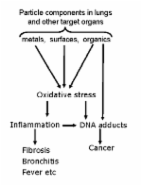
          Particle exposure levels were up to 1 mg/m 3 at the non-commercial airfield. Particulate matter from the non-commercial airfield air consisted of primary and aggregated soot particles, whereas commercial airport sampling resulted in a more heterogeneous mixture of organic compounds including salt, pollen and soot, reflecting the complex occupational exposure at an apron. The particle contents of polycyclic aromatic hydrocarbons and metals were similar to the content in NIST2975. Mice were exposed to doses 6, 18 and 54 μg alongside carbon black (Printex 90) and NIST2975 and euthanized after 1, 28 or 90 days. Dose-dependent increases in total number of cells, neutrophils, and eosinophils in bronchoalveolar lavage fluid were observed on day 1 post-exposure for all particles. Lymphocytes were increased for all four particle types on 28 days post-exposure as well as for neutrophil influx for jet engine particles and carbon black nanoparticles. Increased Saa3 mRNA levels in lung tissue and increased SAA3 protein levels in plasma were observed on day 1 post-exposure. Increased levels of DNA strand breaks in bronchoalveolar lavage cells and liver tissue were observed for both particles, at single dose levels across doses and time points.

          Conclusions

          Pulmonary exposure of mice to particles collected at two airports induced acute phase response, inflammation, and genotoxicity similar to standard diesel exhaust particles and carbon black nanoparticles, suggesting similar physicochemical properties and toxicity of jet engine particles and diesel exhaust particles. Given this resemblance as well as the dose-response relationship between diesel exhaust exposure and lung cancer, occupational exposure to jet engine emissions at the two airports should be minimized.

          Electronic supplementary material

          The online version of this article (10.1186/s12989-019-0305-5) contains supplementary material, which is available to authorized users.

          Related collections

          Most cited references63

          • Record: found
          • Abstract: found
          • Article: not found

          Acute inflammatory responses in the airways and peripheral blood after short-term exposure to diesel exhaust in healthy human volunteers.

          Several epidemiologic studies have demonstrated a consistent association between levels of particulate matter (PM) in the ambient air with increases in cardiovascular and respiratory mortality and morbidity. Diesel exhaust (DE), in addition to generating other pollutants, is a major contributor to PM pollution in most places in the world. Although the epidemiologic evidence is strong, there are as yet no established biological mechanisms to explain the toxicity of PM in humans. To determine the impact of DE on human airways, we exposed 15 healthy human volunteers to air and diluted DE under controlled conditions for 1 h with intermittent exercise. Lung functions were measured before and after each exposure. Blood sampling and bronchoscopy were performed 6 h after each exposure to obtain airway lavages and endobronchial biopsies. While standard lung function measures did not change following DE exposure, there was a significant increase in neutrophils and B lymphocytes in airway lavage, along with increases in histamine and fibronectin. The bronchial biopsies obtained 6 h after DE exposure showed a significant increase in neutrophils, mast cells, CD4+ and CD8+ T lymphocytes along with upregulation of the endothelial adhesion molecules ICAM-1 and VCAM-1, with increases in the numbers of LFA-1+ cells in the bronchial tissue. Significant increases in neutrophils and platelets were observed in peripheral blood following DE exposure. This study demonstrates that at high ambient concentrations, acute short-term DE exposure produces a well-defined and marked systemic and pulmonary inflammatory response in healthy human volunteers, which is underestimated by standard lung function measurements.
            Bookmark
            • Record: found
            • Abstract: found
            • Article: found
            Is Open Access

            Combustion-derived nanoparticles: A review of their toxicology following inhalation exposure

            This review considers the molecular toxicology of combustion-derived nanoparticles (CDNP) following inhalation exposure. CDNP originate from a number of sources and in this review we consider diesel soot, welding fume, carbon black and coal fly ash. A substantial literature demonstrates that these pose a hazard to the lungs through their potential to cause oxidative stress, inflammation and cancer; they also have the potential to redistribute to other organs following pulmonary deposition. These different CDNP show considerable heterogeneity in composition and solubility, meaning that oxidative stress may originate from different components depending on the particle under consideration. Key CDNP-associated properties of large surface area and the presence of metals and organics all have the potential to produce oxidative stress. CDNP may also exert genotoxic effects, depending on their composition. CDNP and their components also have the potential to translocate to the brain and also the blood, and thereby reach other targets such as the cardiovascular system, spleen and liver. CDNP therefore can be seen as a group of particulate toxins unified by a common mechanism of injury and properties of translocation which have the potential to mediate a range of adverse effects in the lungs and other organs and warrant further research.
              Bookmark
              • Record: found
              • Abstract: found
              • Article: found
              Is Open Access

              Lung inflammation and genotoxicity following pulmonary exposure to nanoparticles in ApoE-/- mice

              Background The toxic and inflammatory potential of 5 different types of nanoparticles were studied in a sensitive model for pulmonary effects in apolipoprotein E knockout mice (ApoE-/-). We studied the effects instillation or inhalation Printex 90 of carbon black (CB) and compared CB instillation in ApoE-/- and C57 mice. Three and 24 h after pulmonary exposure, inflammation was assessed by mRNA levels of cytokines in lung tissue, cell composition, genotoxicity, protein and lactate dehydrogenase activity in broncho-alveolar lavage (BAL) fluid. Results Firstly, we found that intratracheal instillation of CB caused far more pulmonary toxicity in ApoE-/- mice than in C57 mice. Secondly, we showed that instillation of CB was more toxic than inhalation of a presumed similar dose with respect to inflammation in the lungs of ApoE-/- mice. Thirdly, we compared effects of instillation in ApoE-/- mice of three carbonaceous particles; CB, fullerenes C60 (C60) and single walled carbon nanotubes (SWCNT) as well as gold particles and quantum dots (QDs). Characterization of the instillation media revealed that all particles were delivered as agglomerates and aggregates. Significant increases in Il-6, Mip-2 and Mcp-1 mRNA were detected in lung tissue, 3 h and 24 h following instillation of SWCNT, CB and QDs. DNA damage in BAL cells, the fraction of neutrophils in BAL cells and protein in BAL fluid increased statistically significantly. Gold and C60 particles caused much weaker inflammatory responses. Conclusion Our data suggest that ApoE-/- model is sensitive for evaluating particle induced inflammation. Overall QDs had greatest effects followed by CB and SWCNT with C60 and gold being least inflammatory and DNA-damaging. However the gold was used at a much lower mass dose than the other particles. The strong effects of QDs were likely due to Cd release. The surface area of the instilled dose correlated well the inflammatory response for low toxicity particles.
                Bookmark

                Author and article information

                Contributors
                kmb@nfa.dk
                abbl@dtu.dk , xabl@nfa.dk
                jok@nrcwe.dk
                ik@force.dk
                trb@nfa.dk
                nib@nrcwe.dk
                kili@dtu.dk
                miikka.dalmaso@tut.fi
                oskari.kangasniemi@tut.fi
                mikko.poikkimaki@tut.fi
                kals@food.dtu.dk
                pac@nfa.dk
                henrik.wolff@ttl.fi
                kaj@nfa.dk
                ats@nfa.dk
                ubv@nrcwe.dk
                Journal
                Part Fibre Toxicol
                Part Fibre Toxicol
                Particle and Fibre Toxicology
                BioMed Central (London )
                1743-8977
                11 June 2019
                11 June 2019
                2019
                : 16
                : 23
                Affiliations
                [1 ]ISNI 0000 0000 9531 3915, GRID grid.418079.3, National Research Centre for the Working Environment, ; Lersø Parkallé 105, DK-2100 Copenhagen, Denmark
                [2 ]ISNI 0000 0001 2181 8870, GRID grid.5170.3, National Centre for Nano Fabrication and Characterization, , Technical University of Denmark, ; Fysikvej, Building 307, DK-2800 Kgs Lyngby, Denmark
                [3 ]ISNI 0000 0004 0606 8858, GRID grid.7320.6, FORCE Technology, ; Park Allé 345, 2605 Brøndby, Denmark
                [4 ]ISNI 0000 0001 2314 6254, GRID grid.502801.e, Aerosol Physics, Laboratory of Physics, Faculty of Natural Sciences, , Tampere University of Technology, ; PO Box 527, FI-33101 Tampere, Finland
                [5 ]ISNI 0000 0001 2181 8870, GRID grid.5170.3, National Food Institute, Research Group for Nano-Bio Science, , Technical University of Denmark, ; Kemitorvet 201, DK-2800 Kgs Lyngby, Denmark
                [6 ]ISNI 0000 0004 0410 5926, GRID grid.6975.d, Finnish Institute of Occupational Health, ; P.O. Box 40, FI-00032, Työterveyslaitos, Helsinki, Finland
                [7 ]ISNI 0000 0001 2181 8870, GRID grid.5170.3, Department of Health Technology, , Technical University of Denmark, ; DK-2800 Kgs Lyngby, Denmark
                Author information
                http://orcid.org/0000-0001-6807-1524
                Article
                305
                10.1186/s12989-019-0305-5
                6558896
                31182125
                38ad83ff-a8c5-4b2c-a379-d4a44e407994
                © The Author(s). 2019

                Open AccessThis article is distributed under the terms of the Creative Commons Attribution 4.0 International License ( http://creativecommons.org/licenses/by/4.0/), which permits unrestricted use, distribution, and reproduction in any medium, provided you give appropriate credit to the original author(s) and the source, provide a link to the Creative Commons license, and indicate if changes were made. The Creative Commons Public Domain Dedication waiver ( http://creativecommons.org/publicdomain/zero/1.0/) applies to the data made available in this article, unless otherwise stated.

                History
                : 30 November 2018
                : 16 May 2019
                Categories
                Research
                Custom metadata
                © The Author(s) 2019

                Toxicology
                airport,exposure risk,jet engine emission,jet engine particle,occupational exposure
                Toxicology
                airport, exposure risk, jet engine emission, jet engine particle, occupational exposure

                Comments

                Comment on this article