2
views
0
recommends
+1 Recommend
0 collections
    0
    shares
      • Record: found
      • Abstract: found
      • Article: found
      Is Open Access

      Experimental Animal Model Systems for Understanding Salivary Secretory Disorders

      review-article

      Read this article at

      Bookmark
          There is no author summary for this article yet. Authors can add summaries to their articles on ScienceOpen to make them more accessible to a non-specialist audience.

          Abstract

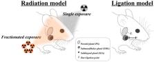
          Salivary secretory disorders are life-disrupting pathologic conditions with a high prevalence, especially in the geriatric population. Both patients and clinicians frequently feel helpless and get frustrated by the currently available therapeutic strategies, which consist mainly of palliative managements. Accordingly, to unravel the underlying mechanisms and to develop effective and curative strategies, several animal models have been developed and introduced. Experimental findings from these models have contributed to answer biological and biomedical questions. This review aims to provide various methodological considerations used for the examination of pathological fundamentals in salivary disorders using animal models and to summarize the obtained findings. The information provided in this review could provide plausible solutions for overcoming salivary disorders and also suggest purpose-specific experimental animal systems.

          Related collections

          Most cited references122

          • Record: found
          • Abstract: found
          • Article: not found

          Global Cancer Statistics 2018: GLOBOCAN Estimates of Incidence and Mortality Worldwide for 36 Cancers in 185 Countries

          This article provides a status report on the global burden of cancer worldwide using the GLOBOCAN 2018 estimates of cancer incidence and mortality produced by the International Agency for Research on Cancer, with a focus on geographic variability across 20 world regions. There will be an estimated 18.1 million new cancer cases (17.0 million excluding nonmelanoma skin cancer) and 9.6 million cancer deaths (9.5 million excluding nonmelanoma skin cancer) in 2018. In both sexes combined, lung cancer is the most commonly diagnosed cancer (11.6% of the total cases) and the leading cause of cancer death (18.4% of the total cancer deaths), closely followed by female breast cancer (11.6%), prostate cancer (7.1%), and colorectal cancer (6.1%) for incidence and colorectal cancer (9.2%), stomach cancer (8.2%), and liver cancer (8.2%) for mortality. Lung cancer is the most frequent cancer and the leading cause of cancer death among males, followed by prostate and colorectal cancer (for incidence) and liver and stomach cancer (for mortality). Among females, breast cancer is the most commonly diagnosed cancer and the leading cause of cancer death, followed by colorectal and lung cancer (for incidence), and vice versa (for mortality); cervical cancer ranks fourth for both incidence and mortality. The most frequently diagnosed cancer and the leading cause of cancer death, however, substantially vary across countries and within each country depending on the degree of economic development and associated social and life style factors. It is noteworthy that high-quality cancer registry data, the basis for planning and implementing evidence-based cancer control programs, are not available in most low- and middle-income countries. The Global Initiative for Cancer Registry Development is an international partnership that supports better estimation, as well as the collection and use of local data, to prioritize and evaluate national cancer control efforts. CA: A Cancer Journal for Clinicians 2018;0:1-31. © 2018 American Cancer Society.
            Bookmark
            • Record: found
            • Abstract: found
            • Article: not found

            On the mechanism of salivary gland radiosensitivity.

            To contribute to the understanding of the enigmatic radiosensitivity of the salivary glands by analysis of appropriate literature, especially with respect to mechanisms of action of early radiation damage, and to supply information on the possibilities of amelioration of radiation damage to the salivary glands after radiotherapy of head-and-neck cancer. Selected published data on the mechanism of salivary gland radiosensitivity and radioprotection were studied and analyzed. From a classical point of view, the salivary glands should not respond as rapidly to radiation as they appear to do. Next to the suggestion of massive apoptosis, the leakage of granules and subsequent lysis of acinar cells was suggested to be responsible for the acute radiation-induced function loss of the salivary glands. The main problem with these hypotheses is that recently performed assays show no cell loss during the first days after irradiation, while saliva flow is dramatically diminished. The water secretion is selectively hampered during the first days after single-dose irradiation. Literature is discussed that shows that the compromised cells suffer selective radiation damage to the plasma membrane, disturbing signal transduction primarily affecting watery secretion. Although the cellular composition of the submandibular gland and the parotid gland are different, the damage response is very alike. The acute radiation-induced function loss in both salivary glands can be ameliorated by prophylactic treatment with specific receptor agonists. The most probable mechanism of action, explaining the enigmatic high radiosensitivity for early effects, is selective radiation damage to the plasma membrane of the secretory cells, disturbing muscarinic receptor stimulated watery secretion. Later damage is mainly due to classical mitotic cell death of progenitor cells, leading to a hampered replacement capacity of the gland for secretory cells, but is also caused by damage to the extracellular environment, preventing proper cell functioning.
              Bookmark
              • Record: found
              • Abstract: found
              • Article: not found

              The functions of human saliva: A review sponsored by the World Workshop on Oral Medicine VI.

              This narrative review of the functions of saliva was conducted in the PubMed, Embase and Web of Science databases. Additional references relevant to the topic were used, as our key words did not generate references which covered all known functions of saliva. These functions include maintaining a moist oral mucosa which is less susceptible to abrasion, and removal of micro-organisms, desquamated epithelial cells, leucocytes and food debris by swallowing. The mucins form a slimy coating on all surfaces in the mouth and act as a lubricant during such processes as mastication, formation of a food bolus, swallowing and speaking. Saliva provides the fluid in which solid tastants may dissolve and distributes tastants around the mouth to the locations of the taste buds. The hypotonic unstimulated saliva facilitates taste recognition. Salivary amylase is involved in digestion of starches. Saliva acts as a buffer to protect oral, pharyngeal and oesophageal mucosae from orally ingested acid or acid regurgitated from the stomach. Saliva protects the teeth against acid by contributing to the acquired enamel pellicle, which forms a renewable lubricant between opposing tooth surfaces, by being supersaturated with respect to tooth mineral, by containing bicarbonate as a buffer and urea and by facilitating clearance of acidic materials from the mouth. Saliva contains many antibacterial, antiviral and antifungal agents which modulate the oral microbial flora in different ways. Saliva also facilitates the healing of oral wounds. Clearly, saliva has many functions which are needed for proper protection and functioning of the human body.
                Bookmark

                Author and article information

                Journal
                Int J Mol Sci
                Int J Mol Sci
                ijms
                International Journal of Molecular Sciences
                MDPI
                1422-0067
                10 November 2020
                November 2020
                : 21
                : 22
                : 8423
                Affiliations
                [1 ]Department of Dental Hygiene, College of Health Science, Gachon University, Incheon 21936, Korea; hoho6434@ 123456gachon.ac.kr
                [2 ]Department of Oral and Maxillofacial Radiology, School of Dentistry, IHBR, Kyungpook National University, 2177, Dalgubeol-daero, Jung-gu, Daegu 41940, Korea; chan@ 123456knu.ac.kr
                [3 ]Department of Biochemistry, School of Dentistry, IHBR, Kyungpook National University, 2177, Dalgubeol-daero, Jung-gu, Daegu 41940, Korea; jykim91@ 123456knu.ac.kr
                [4 ]Department of Oral Medicine, School of Dentistry, IHBR, Kyungpook National University, 2177, Dalgubeol-daero, Jung-gu, Daegu 41940, Korea
                Author notes
                [* ]Correspondence: widenmy@ 123456knu.ac.kr ; Tel.: +82-53-600-7322; Fax: +82-53-426-2195
                Author information
                https://orcid.org/0000-0002-6752-5683
                Article
                ijms-21-08423
                10.3390/ijms21228423
                7696548
                33182571
                390f8fac-e092-441c-a23a-3c1eaedef73c
                © 2020 by the authors.

                Licensee MDPI, Basel, Switzerland. This article is an open access article distributed under the terms and conditions of the Creative Commons Attribution (CC BY) license ( http://creativecommons.org/licenses/by/4.0/).

                History
                : 19 October 2020
                : 05 November 2020
                Categories
                Review

                Molecular biology
                animal experimental model,salivary dysfunction,salivary gland,therapeutic strategies

                Comments

                Comment on this article