44
views
0
recommends
+1 Recommend
0 collections
    0
    shares
      • Record: found
      • Abstract: found
      • Article: found
      Is Open Access

      Myocardial scarring on cardiovascular magnetic resonance in asymptomatic or minimally symptomatic patients with “pure” apical hypertrophic cardiomyopathy

      research-article

      Read this article at

      Bookmark
          There is no author summary for this article yet. Authors can add summaries to their articles on ScienceOpen to make them more accessible to a non-specialist audience.

          Abstract

          Background

          Late gadolinium enhancement (LGE) cardiovascular magnetic resonance (CMR) enables state-of-the-art in vivo evaluations of myocardial fibrosis. Although LGE patterns have been well described in asymmetrical septal hypertrophy, conflicting results have been reported regarding the characteristics of LGE in apical hypertrophic cardiomyopathy (ApHCM). This study was undertaken to determine 1) the frequency and distribution of LGE and 2) its prognostic implication in ApHCM.

          Methods

          Forty patients with asymptomatic or minimally symptomatic pure ApHCM (age, 60.2 ± 10.4 years, 31 men) were prospectively enrolled. LGE images were acquired using the inversion recovery segmented spoiled-gradient echo and phase-sensitive inversion recovery sequence, and analyzed using a 17-segment model. Summing the planimetered LGE areas in all short axis slices yielded the total volume of late enhancement, which was subsequently presented as a proportion of total LV myocardium (% LGE).

          Results

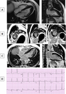
          Mean maximal apical wall thickness was 17.9±2.3mm, and mean left ventricular (LV) ejection fraction was 67.7 ± 8.0%. All but one patient presented with electrocardiographic negative T wave inversion in anterolateral leads, with a mean maximum negative T wave of 7.2 ± 4.7mm. Nine patients (22.5%) had giant negative T waves, defined as the amplitude of ≥10mm, in electrocardiogram. LGE was detected in 130 segments of 30 patients (75.0%), occupying 4.9 ± 5.5% of LV myocardium. LGE was mainly detected at the junction between left and right ventricles in 12 (30%) and at the apex in 28 (70%), although LGE-positive areas were widely distributed, and not limited to the apex. Focal LGE at the non-hypertrophic LV segments was found in some ApHCM patients, even without LGE of hypertrophied apical segments. Over the 2-year follow-up, there was no one achieving the study end-point, defined as all-cause death, sudden cardiac death and hospitalization for heart failure.

          Conclusions

          LGE was frequently observed not only in the thickened apex of the heart but also in other LV segments, irrespective of the presence or absence of hypertrophy. The simple presence of LGE on CMR was not representative of adverse prognosis in this population.

          Related collections

          Most cited references22

          • Record: found
          • Abstract: found
          • Article: not found

          Hypertrophic cardiomyopathy: a systematic review.

          Throughout the past 40 years, a vast and sometimes contradictory literature has accumulated regarding hypertrophic cardiomyopathy (HCM), a genetic cardiac disease caused by a variety of mutations in genes encoding sarcomeric proteins and characterized by a broad and expanding clinical spectrum. To clarify and summarize the relevant clinical issues and to profile rapidly evolving concepts regarding HCM. Systematic analysis of the relevant HCM literature, accessed through MEDLINE (1966-2000), bibliographies, and interactions with investigators. Diverse information was assimilated into a rigorous and objective contemporary description of HCM, affording greatest weight to prospective, controlled, and evidence-based studies. Hypertrophic cardiomyopathy is a relatively common genetic cardiac disease (1:500 in the general population) that is heterogeneous with respect to disease-causing mutations, presentation, prognosis, and treatment strategies. Visibility attached to HCM relates largely to its recognition as the most common cause of sudden death in the young (including competitive athletes). Clinical diagnosis is by 2-dimensional echocardiographic identification of otherwise unexplained left ventricular wall thickening in the presence of a nondilated cavity. Overall, HCM confers an annual mortality rate of about 1% and in most patients is compatible with little or no disability and normal life expectancy. Subsets with higher mortality or morbidity are linked to the complications of sudden death, progressive heart failure, and atrial fibrillation with embolic stroke. Treatment strategies depend on appropriate patient selection, including drug treatment for exertional dyspnea (beta-blockers, verapamil, disopyramide) and the septal myotomy-myectomy operation, which is the standard of care for severe refractory symptoms associated with marked outflow obstruction; alcohol septal ablation and pacing are alternatives to surgery for selected patients. High-risk patients may be treated effectively for sudden death prevention with the implantable cardioverter-defibrillator. Substantial understanding has evolved regarding the epidemiology and clinical course of HCM, as well as novel treatment strategies that may alter its natural history. An appreciation that HCM, although an important cause of death and disability at all ages, does not invariably convey ominous prognosis and is compatible with normal longevity should dictate a large measure of reassurance for many patients.
            Bookmark
            • Record: found
            • Abstract: found
            • Article: not found

            Delayed enhancement cardiovascular magnetic resonance assessment of non-ischaemic cardiomyopathies.

            Non-ischaemic cardiomyopathies (NICMs) are chronic, progressive myocardial diseases with distinct patterns of morphological, functional, and electrophysiological changes. In the setting of cardiomyopathy (CM), determining the exact aetiology is important because the aetiology is directly related to treatment and patient survival. Determining the exact aetiology, however, can be difficult using currently available imaging techniques, such as echocardiography, radionuclide imaging or X-ray coronary angiography, since overlap of features between CMs may be encountered. Cardiovascular magnetic resonance (CMR) imaging has recently emerged as a new non-invasive imaging modality capable of providing high-resolution images of the heart in any desired plane. Delayed contrast enhanced CMR (DE-CMR) can be used for non-invasive tissue characterization and may hold promise in differentiating ischaemic from NICMs, as the typical pattern of hyperenhancement can be classified as 'ischaemic-type' or 'non-ischaemic type' on the basis of pathophysiology of ischaemia. This article reviews the potential of DE-CMR to distinguish between ischaemic and NICM as well as to differentiate non-ischaemic aetiologies. Rather than simply describing various hyperenhancement patterns that may occur in different disease states, our goal will be (i) to provide an overall imaging approach for the diagnosis of CM and (ii) to demonstrate how this approach is based on the underlying relationships between contrast enhancement and myocardial pathophysiology.
              Bookmark
              • Record: found
              • Abstract: found
              • Article: not found

              The histologic basis of late gadolinium enhancement cardiovascular magnetic resonance in hypertrophic cardiomyopathy.

              We sought to identify the histologic basis of myocardial late gadolinium enhancement cardiovascular magnetic resonance (CMR) in hypertrophic cardiomyopathy (HCM). The histologic basis of late gadolinium CMR in patients with HCM is unknown. A 28-year-old male patient with HCM and heart failure underwent late gadolinium enhancement CMR and, 49 days later, heart transplantation. The explanted heart was examined histologically for the extent of collagen and disarray, and the results were compared with a previous in vivo CMR scan. Overall, 19% of the myocardium was collagen, but the amount per segment varied widely (SD +/- 19, range 0% to 71%). Both disarray and collagen were more likely to be found in the mesocardium than in the endo- or epicardium. There was a significant relationship between the extent of late gadolinium enhancement and collagen (r = 0.7, p 15% collagen were more likely to have late gadolinium enhancement. Regional wall motion was inversely related to the extent of myocardial collagen and late gadolinium enhancement but not disarray (p = 0.0003, 0.04, and NS, respectively). In this patient with HCM and heart failure, regions of myocardial late gadolinium enhancement by CMR represented regions of increased myocardial collagen but not disarray.
                Bookmark

                Author and article information

                Journal
                J Cardiovasc Magn Reson
                J Cardiovasc Magn Reson
                Journal of Cardiovascular Magnetic Resonance
                BioMed Central
                1097-6647
                1532-429X
                2012
                28 July 2012
                : 14
                : 1
                : 52
                Affiliations
                [1 ]Division of Cardiology, Department of Internal Medicine, Cardiovascular Center, Seoul National University College of Medicine, Seoul National University Hospital, 28 Yongon-dong, Chongno-gu, Seoul, 110-744, South Korea
                [2 ]Department of Radiology, Seoul National University College of Medicine, Seoul National University Hospital, 28 Yongon-dong, Chongno-gu, Seoul, 110-744, South Korea
                Article
                1532-429X-14-52
                10.1186/1532-429X-14-52
                3419125
                22839526
                39f2f24b-bca6-4cc6-9fe2-03e4e290ade6
                Copyright ©2012 Kim et al.; licensee BioMed Central Ltd.

                This is an Open Access article distributed under the terms of the Creative Commons Attribution License ( http://creativecommons.org/licenses/by/2.0), which permits unrestricted use, distribution, and reproduction in any medium, provided the original work is properly cited.

                History
                : 17 February 2012
                : 10 July 2012
                Categories
                Research

                Cardiovascular Medicine
                apical hypertrophic cardiomyopathy,late gadolinium enhancement,cardiovascular magnetic resonance

                Comments

                Comment on this article