35
views
0
recommends
+1 Recommend
0 collections
    0
    shares
      • Record: found
      • Abstract: found
      • Article: not found

      Cre- loxP–mediated Inactivation of the α6A Integrin Splice Variant In Vivo: Evidence for a Specific Functional Role of α6A in Lymphocyte Migration but Not in Heart Development

      research-article

      Read this article at

      ScienceOpenPublisherPMC
      Bookmark
          There is no author summary for this article yet. Authors can add summaries to their articles on ScienceOpen to make them more accessible to a non-specialist audience.

          Abstract

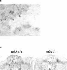
          Two splice variants of the α6 integrin subunit, α6A and α6B, with different cytoplasmic domains, have previously been described. While α6B is expressed throughout the development of the mouse, the expression of α6A begins at 8.5 days post coitum and is initially restricted to the myocardium. Later in ontogeny, α6A is found in various epithelia and in certain cells of the immune system. In this study, we have investigated the function of α6A in vivo by generating knockout mice deficient for this splice variant. The Cre- loxP system of the bacteriophage P1 was used to specifically remove the exon encoding the cytoplasmic domain of α6A in embryonic stem cells, and the deletion resulted in the expression of α6B in all tissues that normally express α6A. We show that α6A−/− mice develop normally and are fertile. The substitution of α6A by α6B does not impair the development and function of the heart, hemidesmosome formation in the epidermis, or keratinocyte migration. Furthermore, T cells differentiated normally in α6A−/− mice. However, the substitution of α6A by α6B leads to a decrease in the migration of lymphocytes through laminin-coated Transwell filters and to a reduction of the number of T cells isolated from the peripheral and mesenteric lymph nodes. Lymphocyte homing to the lymph nodes, which involves various types of integrin–ligand interactions, was not affected in the α6A knockout mice, indicating that the reduced number of lymph node cells could not be directly attributed to defects in lymphocyte trafficking. Nevertheless, the expression of α6A might be necessary for optimal lymphocyte migration on laminin in certain pathological conditions.

          Related collections

          Most cited references68

          • Record: found
          • Abstract: found
          • Article: not found

          Integrins and signal transduction pathways: the road taken.

          Adhesive interactions play critical roles in directing the migration, proliferation, and differentiation of cells; aberrations in such interactions can lead to pathological disorders. These adhesive interactions, mediated by cell surface receptors that bind to ligands on adjacent cells or in the extracellular matrix, also regulate intracellular signal transduction pathways that control adhesion-induced changes in cell physiology. Though the extracellular molecular interactions involving many adhesion receptors have been well characterized, the adhesion-dependent intracellular signaling events that regulate these physiological alterations have only begun to be elucidated. This article will focus on recent advances in our understanding of intracellular signal transduction pathways regulated by the integrin family of adhesion receptors.
            Bookmark
            • Record: found
            • Abstract: found
            • Article: not found

            Beta4 integrin is required for hemidesmosome formation, cell adhesion and cell survival

            The integrin heterodimer alpha 6 beta 4 is expressed in many epithelia and in Schwann cells. In stratified epithelia, alpha 6 beta 4 couple with BPAG1-e and BPAG2 to form hemidesmosomes, attaching externally to laminin and internally to the keratin cytoskeleton. To explore the function of this atypical integrin, and its relation to conventional actin-associated integrins, we targeted the removal of the beta 4 gene in mice. Tissues that express alpha 6 beta 4 are grossly affected. Stratified tissues are devoid of hemidesmosomes, display only a very fragile attachment to the basal lamina, and exhibit signs of degeneration and tissue disorganization. Simple epithelia which express alpha 6 beta 4 are also defective in adherence, even though they do not form hemidesmosomes. In the absence of beta 4, alpha 6 is dramatically downregulated, and other integrins do not appear to compensate for the loss of this heterodimer. These data have important implications for understanding integrin function in cell-substratum adhesion, cell survival and differentiation, and for understanding the role of alpha 6 beta 4 in junctional epidermolysis bullosa, an often lethal human disorder with pathology similar to our mice.
              Bookmark
              • Record: found
              • Abstract: found
              • Article: not found

              Keratinocyte growth factor is required for hair development but not for wound healing.

              Keratinocyte growth factor (KGF), also known as fibroblast growth factor 7 (FGF7), is synthesized by skin fibroblasts. However, its mitogenic activity is on skin keratinocytes, where it is the most potent growth factor identified thus far. To explore KGF's function in vivo, we used embryonic stem cell technology to generate mice lacking KGF. Over time, their fur developed a matted appearance, very similar to that of the rough mouse, whose recessive mutation maps at or near the KGF locus on mouse chromosome 2. In contrast to the recently reported transforming growth factor-alpha (TGF-alpha) and FGF5 knockouts, which showed defects in the follicle outer-root sheath and the hair growth cycle, respectively, the hair defect in the KGF knockout seemed to be restricted to the cells giving rise to the hair shaft. Thus, we have uncovered a third, and at least partially nonoverlapping, growth factor pathway involved in orchestrating hair follicle growth and/or differentiation. Surprisingly, the absence of KGF resulted in no abnormalities in epidermal growth or wound healing. This was true even when we engineered double knockout mice, null for both KGF and TGF-alpha, two factors that are increased dramatically in the normal wound-healing process. Whereas we found no evidence of compensatory changes at the mRNA level of wounded knockout mice, these data imply that the regulation of epidermal growth is complex and involves a number of growth stimulatory factors that go beyond what are thought to be the major paracrine and autocrine growth factors. We suggest that the redundancy in epidermal growth and wound healing is likely to stem from the vitality of these functions to the organism, a feature that is not a consideration for the hair follicle.
                Bookmark

                Author and article information

                Journal
                J Cell Biol
                The Journal of Cell Biology
                The Rockefeller University Press
                0021-9525
                1540-8140
                5 October 1998
                : 143
                : 1
                : 253-266
                Affiliations
                Division of Cell Biology, The Netherlands Cancer Institute, Plesmanlaan 121, 1066 CX Amsterdam, The Netherlands
                Article
                10.1083/jcb.143.1.253
                2132821
                9763436
                39fba4b7-04b5-40df-8731-95bb7950d15a
                Copyright @ 1998
                History
                : 20 April 1998
                : 21 July 1998
                Categories
                Regular Articles

                Cell biology
                integrin,laminin receptor,knockout,migration,lymphocyte
                Cell biology
                integrin, laminin receptor, knockout, migration, lymphocyte

                Comments

                Comment on this article