12
views
0
recommends
+1 Recommend
0 collections
    0
    shares
      • Record: found
      • Abstract: found
      • Article: found
      Is Open Access

      Cognitive Rehabilitation in Bilateral Vestibular Patients: A Computational Perspective

      brief-report

      Read this article at

      Bookmark
          There is no author summary for this article yet. Authors can add summaries to their articles on ScienceOpen to make them more accessible to a non-specialist audience.

          Abstract

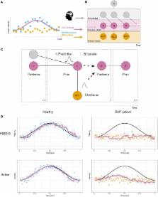
          There is evidence that vestibular sensory processing affects, and is affected by, higher cognitive processes. This is highly relevant from a clinical perspective, where there is evidence for cognitive impairments in patients with peripheral vestibular deficits. The vestibular system performs complex probabilistic computations, and we claim that understanding these is important for investigating interactions between vestibular processing and cognition. Furthermore, this will aid our understanding of patients’ self-motion perception and will provide useful information for clinical interventions. We propose that cognitive training is a promising way to alleviate the debilitating symptoms of patients with complete bilateral vestibular loss (BVP), who often fail to show improvement when relying solely on conventional treatment methods. We present a probabilistic model capable of processing vestibular sensory data during both passive and active self-motion. Crucially, in our model, knowledge from multiple sources, including higher-level cognition, can be used to predict head motion. This is the entry point for cognitive interventions. Despite the loss of sensory input, the processing circuitry in BVP patients is still intact, and they can still perceive self-motion when the movement is self-generated. We provide computer simulations illustrating self-motion perception of BVP patients. Cognitive training may lead to more accurate and confident predictions, which result in decreased weighting of sensory input, and thus improved self-motion perception. Using our model, we show the possible impact of cognitive interventions to help vestibular rehabilitation in patients with BVP.

          Related collections

          Most cited references49

          • Record: found
          • Abstract: found
          • Article: not found

          Object perception as Bayesian inference.

          We perceive the shapes and material properties of objects quickly and reliably despite the complexity and objective ambiguities of natural images. Typical images are highly complex because they consist of many objects embedded in background clutter. Moreover, the image features of an object are extremely variable and ambiguous owing to the effects of projection, occlusion, background clutter, and illumination. The very success of everyday vision implies neural mechanisms, yet to be understood, that discount irrelevant information and organize ambiguous or noisy local image features into objects and surfaces. Recent work in Bayesian theories of visual perception has shown how complexity may be managed and ambiguity resolved through the task-dependent, probabilistic integration of prior object knowledge with image features.
            Bookmark
            • Record: found
            • Abstract: found
            • Article: not found

            Vestibular loss causes hippocampal atrophy and impaired spatial memory in humans.

            The human hippocampal formation plays a crucial role in various aspects of memory processing. Most literature on the human hippocampus stresses its non-spatial memory functions, but older work in rodents and some other species emphasized the role of the hippocampus in spatial learning and memory as well. A few human studies also point to a direct relation between hippocampal size, navigation and spatial memory. Conversely, the importance of the vestibular system for navigation and spatial memory was until now convincingly demonstrated only in animals. Using magnetic resonance imaging volumetry, we found that patients (n = 10) with acquired chronic bilateral vestibular loss (BVL) develop a significant selective atrophy of the hippocampus (16.9% decrease relative to controls). When tested with a virtual variant (on a PC) of the Morris water task these patients exhibited significant spatial memory and navigation deficits that closely matched the pattern of hippocampal atrophy. These spatial memory deficits were not associated with general memory deficits. The current data on BVL patients and bilateral hippocampal atrophy revive the idea that a major--and probably phylogenetically ancient--function of the archicortical hippocampal tissue is still evident in spatial aspects of memory processing for navigation. Furthermore, these data demonstrate for the first time in humans that spatial navigation critically depends on preserved vestibular function, even when the subjects are stationary, e.g. without any actual vestibular or somatosensory stimulation.
              Bookmark
              • Record: found
              • Abstract: found
              • Article: found
              Is Open Access

              Bilateral vestibulopathy: Diagnostic criteria Consensus document of the Classification Committee of the Bárány Society

              This paper describes the diagnostic criteria for bilateral vestibulopathy (BVP) by the Classification Committee of the Bárány Society. The diagnosis of BVP is based on the patient history, bedside examination and laboratory evaluation. Bilateral vestibulopathy is a chronic vestibular syndrome which is characterized by unsteadiness when walking or standing, which worsen in darkness and/or on uneven ground, or during head motion. Additionally, patients may describe head or body movement-induced blurred vision or oscillopsia. There are typically no symptoms while sitting or lying down under static conditions. The diagnosis of BVP requires bilaterally significantly impaired or absent function of the vestibulo-ocular reflex (VOR). This can be diagnosed for the high frequency range of the angular VOR by the head impulse test (HIT), the video-HIT (vHIT) and the scleral coil technique and for the low frequency range by caloric testing. The moderate range can be examined by the sinusoidal or step profile rotational chair test. For the diagnosis of BVP, the horizontal angular VOR gain on both sides should be <0.6 (angular velocity 150–300°/s) and/or the sum of the maximal peak velocities of the slow phase caloric-induced nystagmus for stimulation with warm and cold water on each side <6°/s and/or the horizontal angular VOR gain <0.1 upon sinusoidal stimulation on a rotatory chair (0.1 Hz, Vmax = 50°/sec) and/or a phase lead >68 degrees (time constant of <5 seconds). For the diagnosis of probable BVP the above mentioned symptoms and a bilaterally pathological bedside HIT are required. Complementary tests that may be used but are currently not included in the definition are: a) dynamic visual acuity (a decrease of ≥0.2 logMAR is considered pathological); b) Romberg (indicating a sensory deficit of the vestibular or somatosensory system and therefore not specific); and c) abnormal cervical and ocular vestibular-evoked myogenic potentials for otolith function. At present the scientific basis for further subdivisions into subtypes of BVP is not sufficient to put forward reliable or clinically meaningful definitions. Depending on the affected anatomical structure and frequency range, different subtypes may be better identified in the future: impaired canal function in the low- or high-frequency VOR range only and/or impaired otolith function only; the latter is evidently very rare. Bilateral vestibulopathy is a clinical syndrome and, if known, the etiology (e.g., due to ototoxicity, bilateral Menière’s disease, bilateral vestibular schwannoma) should be added to the diagnosis. Synonyms include bilateral vestibular failure, deficiency, areflexia, hypofunction and loss.
                Bookmark

                Author and article information

                Contributors
                Journal
                Front Neurol
                Front Neurol
                Front. Neurol.
                Frontiers in Neurology
                Frontiers Media S.A.
                1664-2295
                27 April 2018
                2018
                : 9
                : 286
                Affiliations
                [1] 1Department of Psychology, University of Bern , Bern, Switzerland
                [2] 2Center for Cognition, Learning and Memory, University of Bern , Bern, Switzerland
                [3] 3Department of Otorhinolaryngology, Head and Neck Surgery, Inselspital, University Hospital Bern, University of Bern , Bern, Switzerland
                Author notes

                Edited by: Alexander A. Tarnutzer, Universität Zürich, Switzerland

                Reviewed by: W. Pieter Medendorp, Radboud University Nijmegen, Netherlands; Silvia Colnaghi, University of Pavia, Italy

                *Correspondence: Andrew W. Ellis, andrew.ellis@ 123456psy.unibe.ch

                Specialty section: This article was submitted to Neuro-Otology, a section of the journal Frontiers in Neurology

                Article
                10.3389/fneur.2018.00286
                5934854
                29755404
                3aa42406-3e76-4f14-88cc-321de3cb70c8
                Copyright © 2018 Ellis, Schöne, Vibert, Caversaccio and Mast.

                This is an open-access article distributed under the terms of the Creative Commons Attribution License (CC BY). The use, distribution or reproduction in other forums is permitted, provided the original author(s) and the copyright owner are credited and that the original publication in this journal is cited, in accordance with accepted academic practice. No use, distribution or reproduction is permitted which does not comply with these terms.

                History
                : 31 January 2018
                : 13 April 2018
                Page count
                Figures: 2, Tables: 0, Equations: 0, References: 52, Pages: 7, Words: 4962
                Funding
                Funded by: Schweizerischer Nationalfonds zur Förderung der Wissenschaftlichen Forschung 10.13039/501100001711
                Award ID: CR13I1_147164, 100014_162480
                Categories
                Neuroscience
                Perspective

                Neurology
                cognitive training,bilateral vestibulopathy,bilateral vestibular loss,rehabilitation,vestibular cognition,computational modeling,mental imagery,self-motion perception

                Comments

                Comment on this article