8
views
0
recommends
+1 Recommend
0 collections
    0
    shares
      • Record: found
      • Abstract: found
      • Article: found
      Is Open Access

      Effects of Platelet-Rich Plasma on Kidney Regeneration in Gentamicin-Induced Nephrotoxicity

      research-article

      Read this article at

      Bookmark
          There is no author summary for this article yet. Authors can add summaries to their articles on ScienceOpen to make them more accessible to a non-specialist audience.

          Abstract

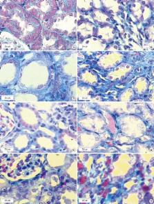
          Platelet-rich plasma (PRP) as a source of growth factors may induce tissue repairing and improve fibrosis. This study aimed to assess the effects of PRP on kidney regeneration and fibrosis in gentamicin (GM)-induced nephrotoxicity rat model by stereological study. Thirty-two male rats were selected. Nephrotoxicity was induced in animals by administration of GM (80 mg/kg/daily, intraperitoneally [IP], 8 day) and animals were treated by PRP (100 µL, intra-cortical injection using surgical microscopy, single dose). Blood samples were collected for determine blood urea nitrogen (BUN) and creatinine (Cr) before and after PRP therapy. At the end of experiment, right kidneys were sectioned by Isotropic Uniform Random (IUR) method and stained with H & E and Masson’s Trichrome. The stereological methods were used for estimating the changes in different structures of kidney. PRP increased the number of epithelial cells in convoluted tubules, and decreased the volume of connective tissue, renal corpuscles and glomeruli in GM-treated animals ( P < 0.05). Our findings indicate that PRP had beneficial effects on proliferation of epithelial cells in convoluted tubules and ameliorated GM-induced fibrosis.

          Graphical Abstract

          Related collections

          Most cited references23

          • Record: found
          • Abstract: found
          • Article: not found

          The role of growth factors in cartilage repair.

          Full-thickness chondral defects and early osteoarthritis continue to present major challenges for the patient and the orthopaedic surgeon as a result of the limited healing potential of articular cartilage. The use of bioactive growth factors is under consideration as a potential therapy to enhance healing of chondral injuries and modify the arthritic disease process. We reviewed the role of growth factors in articular cartilage repair and identified specific growth factors and combinations of growth factors that have the capacity to improve cartilage regeneration. Additionally, we discuss the potential use of platelet-rich plasma, autologous-conditioned serum, and bone marrow concentrate preparations as methods of combined growth factor delivery. A PubMed search was performed using key words cartilage or chondrocyte alone and in combination with growth factor. The search was open for original manuscripts and review papers and open for all dates. From these searches we selected manuscripts investigating the effects of growth factors on extracellular matrix synthesis and excluded those investigating molecular mechanisms of action. By modulating the local microenvironment, the anabolic and anticatabolic effects of a variety of growth factors have demonstrated potential in both in vitro and animal studies of cartilage injury and repair. Members of the transforming growth factor-β superfamily, fibroblast growth factor family, insulin-like growth factor-I, and platelet-derived growth factor have all been investigated as possible treatment augments in the management of chondral injuries and early arthritis. The application of growth factors in the treatment of local cartilage defects as well as osteoarthritis appears promising; however, further research is needed at both the basic science and clinical levels before routine application.
            Bookmark
            • Record: found
            • Abstract: found
            • Article: not found

            Positive effect of an autologous platelet concentrate in lateral epicondylitis in a double-blind randomized controlled trial: platelet-rich plasma versus corticosteroid injection with a 1-year follow-up.

            Platelet-rich plasma (PRP) has shown to be a general stimulation for repair. Purpose To determine the effectiveness of PRP compared with corticosteroid injections in patients with chronic lateral epicondylitis. Randomized controlled trial; Level of evidence, 1. The trial was conducted in 2 teaching hospitals in the Netherlands. One hundred patients with chronic lateral epicondylitis were randomly assigned in the PRP group (n = 51) or the corticosteroid group (n = 49). A central computer system carried out randomization and allocation to the trial group. Patients were randomized to receive either a corticosteroid injection or an autologous platelet concentrate injection through a peppering technique. The primary analysis included visual analog scores and DASH Outcome Measure scores (DASH: Disabilities of the Arm, Shoulder, and Hand). Successful treatment was defined as more than a 25% reduction in visual analog score or DASH score without a reintervention after 1 year. The results showed that, according to the visual analog scores, 24 of the 49 patients (49%) in the corticosteroid group and 37 of the 51 patients (73%) in the PRP group were successful, which was significantly different (P <.001). Furthermore, according to the DASH scores, 25 of the 49 patients (51%) in the corticosteroid group and 37 of the 51 patients (73%) in the PRP group were successful, which was also significantly different (P = .005). The corticosteroid group was better initially and then declined, whereas the PRP group progressively improved. Treatment of patients with chronic lateral epicondylitis with PRP reduces pain and significantly increases function, exceeding the effect of corticosteroid injection. Future decisions for application of the PRP for lateral epicondylitis should be confirmed by further follow-up from this trial and should take into account possible costs and harms as well as benefits.
              Bookmark
              • Record: found
              • Abstract: found
              • Article: not found

              TGF-beta signaling in renal disease.

              Since discovery over a decade ago of a role for the cytokine TGF-beta as key mediator of glomerular and tubulointerstitial pathobiology in chronic kidney diseases, studies of TGF-beta signaling in the kidney have focused on the molecular biology of fibrogenesis. In recent years, glomerular and tubular epithelial cell apoptosis and cellular transdifferentiation have been proposed as putative primary pathomechanisms that may underlie progression of renal disease. This review describes evidence in support of nonlinear models and functional roles of TGF-beta signaling in mediating apoptosis and epithelial-to-mesenchymal transdifferentiation (EMT) in chronic progressive renal disease. Emphasis is placed on cell context-dependent models of TGF-beta signaling providing a conceptual framework to consolidate seemingly distinct pathomechanisms of progression of glomerular and tubulointerstitial disease.
                Bookmark

                Author and article information

                Journal
                J Korean Med Sci
                J. Korean Med. Sci
                JKMS
                Journal of Korean Medical Science
                The Korean Academy of Medical Sciences
                1011-8934
                1598-6357
                January 2017
                08 November 2016
                : 32
                : 1
                : 13-21
                Affiliations
                [1 ]Department of Anatomy, Shiraz University of Medical Sciences, Shiraz, Iran.
                [2 ]Histomorphometry and Stereology Research Center, Shiraz University of Medical Sciences, Shiraz, Iran.
                Author notes
                Address for Correspondence: Farzaneh Dehghani, PhD. Department of Anatomy, Shiraz University of Medical Sciences, Zand Ave, Shiraz 71934, Iran. dehghanf@ 123456sums.ac.ir
                Author information
                http://orcid.org/0000-0002-5107-5569
                http://orcid.org/0000-0002-8425-8871
                http://orcid.org/0000-0002-7160-5088
                http://orcid.org/0000-0002-6635-2717
                http://orcid.org/0000-0003-2217-5672
                Article
                10.3346/jkms.2017.32.1.13
                5143284
                27914126
                3b2dc7df-fede-40c3-a45b-4d4ab3ef5700
                © 2017 The Korean Academy of Medical Sciences.

                This is an Open Access article distributed under the terms of the Creative Commons Attribution Non-Commercial License ( http://creativecommons.org/licenses/by-nc/4.0/) which permits unrestricted non-commercial use, distribution, and reproduction in any medium, provided the original work is properly cited.

                History
                : 14 April 2016
                : 20 August 2016
                Funding
                Funded by: Shiraz University of Medical Sciences, CrossRef http://dx.doi.org/10.13039/501100004320;
                Award ID: 92-6792
                Categories
                Original Article
                Basic Medical Sciences

                Medicine
                fibrosis,kidney,regeneration
                Medicine
                fibrosis, kidney, regeneration

                Comments

                Comment on this article