159
views
0
recommends
+1 Recommend
0 collections
    0
    shares
      • Record: found
      • Abstract: found
      • Article: found
      Is Open Access

      Motion correction in optical coherence tomography volumes on a per A-scan basis using orthogonal scan patterns

      research-article

      Read this article at

      Bookmark
          There is no author summary for this article yet. Authors can add summaries to their articles on ScienceOpen to make them more accessible to a non-specialist audience.

          Abstract

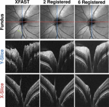
          High speed Optical Coherence Tomography (OCT) has made it possible to rapidly capture densely sampled 3D volume data. One key application is the acquisition of high quality in vivo volumetric data sets of the human retina. Since the volume is acquired in a few seconds, eye movement during the scan process leads to distortion, which limits the accuracy of quantitative measurements using 3D OCT data. In this paper, we present a novel software based method to correct motion artifacts in OCT raster scans. Motion compensation is performed retrospectively using image registration algorithms on the OCT data sets themselves. Multiple, successively acquired volume scans with orthogonal fast scan directions are registered retrospectively in order to estimate and correct eye motion. Registration is performed by optimizing a large scale numerical problem as given by a global objective function using one dense displacement field for each input volume and special regularization based on the time structure of the acquisition process. After optimization, each volume is undistorted and a single merged volume is constructed that has superior signal quality compared to the input volumes. Experiments were performed using 3D OCT data from the macula and optic nerve head acquired with a high-speed ultra-high resolution 850 nm spectral OCT as well as wide field data acquired with a 1050 nm swept source OCT instrument. Evaluation of registration performance and result stability as well as visual inspection shows that the algorithm can correct for motion in all three dimensions and on a per A-scan basis. Corrected volumes do not show visible motion artifacts. In addition, merging multiple motion corrected and registered volumes leads to improved signal quality. These results demonstrate that motion correction and merging improves image quality and should also improve morphometric measurement accuracy from volumetric OCT data.

          Related collections

          Most cited references19

          • Record: found
          • Abstract: found
          • Article: not found

          Optical coherence tomography.

          A technique called optical coherence tomography (OCT) has been developed for noninvasive cross-sectional imaging in biological systems. OCT uses low-coherence interferometry to produce a two-dimensional image of optical scattering from internal tissue microstructures in a way that is analogous to ultrasonic pulse-echo imaging. OCT has longitudinal and lateral spatial resolutions of a few micrometers and can detect reflected signals as small as approximately 10(-10) of the incident optical power. Tomographic imaging is demonstrated in vitro in the peripapillary area of the retina and in the coronary artery, two clinically relevant examples that are representative of transparent and turbid media, respectively.
            Bookmark
            • Record: found
            • Abstract: not found
            • Article: not found

            A new approach to variable metric algorithms

              Bookmark
              • Record: found
              • Abstract: found
              • Article: not found

              In vivo retinal imaging by optical coherence tomography.

              We describe what are to our knowledge the first in vivo measurements of human retinal structure with optical coherence tomography. These images represent the highest depth resolution in vivo retinal images to date. The tomographic system, image-processing techniques, and examples of high-resolution tomographs and their clinical relevance are discussed.
                Bookmark

                Author and article information

                Journal
                Biomed Opt Express
                Biomed Opt Express
                BOE
                Biomedical Optics Express
                Optical Society of America
                2156-7085
                03 May 2012
                01 June 2012
                03 May 2012
                : 3
                : 6
                : 1182-1199
                Affiliations
                [1 ]Pattern Recognition Lab, University Erlangen-Nuremberg, Erlangen, Germany
                [2 ]School of Advanced Optical Technologies (SAOT), University Erlangen-Nuremberg, Germany
                [3 ]Dept of Electrical Engineering and Computer Science and Research Laboratory of Electronics, Massachusetts Inst. of Technology, Cambridge, MA, USA
                [4 ]Advanced Imaging Group, Thorlabs, Inc., Newton, NJ 07860, USA
                Author notes
                Article
                164364
                10.1364/BOE.3.001182
                3370961
                22741067
                3d42f594-da69-4ab8-a83b-bd095ff4dca5
                ©2012 Optical Society of America

                This is an open-access article distributed under the terms of the Creative Commons Attribution-Noncommercial-No Derivative Works 3.0 Unported License, which permits download and redistribution, provided that the original work is properly cited. This license restricts the article from being modified or used commercially.

                History
                : 9 March 2012
                : 23 April 2012
                : 25 April 2012
                Funding
                Funded by: National Institutes of Health
                Award ID: R01-EY011289-25
                Award ID: R01-EY013178-11
                Award ID: R01-EY013516-09
                Award ID: R01-EY019029-03
                Award ID: R01-HL095717-03
                Award ID: R01-NS057476-05
                Funded by: Air Force Office of Scientific Research
                Award ID: FA9550-10-1-0063
                Funded by: Medical Free Electron Laser Program
                Award ID: FA9550-10-1-0551
                Funded by: German Research Foundation (DFG)
                Categories
                Optical Coherence Tomography
                Custom metadata
                True
                0

                Vision sciences
                (170.4500) optical coherence tomography,(100.5010) pattern recognition,(170.4470) ophthalmology,(100.2980) image enhancement

                Comments

                Comment on this article