33
views
0
recommends
+1 Recommend
0 collections
    0
    shares
      • Record: found
      • Abstract: found
      • Article: found
      Is Open Access

      Successful tumour necrosis factor (TNF) blocking therapy suppresses oxidative stress and hypoxia-induced mitochondrial mutagenesis in inflammatory arthritis

      research-article

      Read this article at

      Bookmark
          There is no author summary for this article yet. Authors can add summaries to their articles on ScienceOpen to make them more accessible to a non-specialist audience.

          Abstract

          Introduction

          To examine the effects of tumour necrosis factor (TNF) blocking therapy on the levels of early mitochondrial genome alterations and oxidative stress.

          Methods

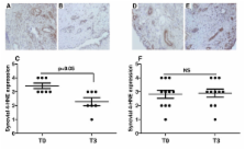
          Eighteen inflammatory arthritis patients underwent synovial tissue oxygen (tpO 2) measurements and clinical assessment of disease activity (DAS28-CRP) at baseline (T0) and three months (T3) after starting biologic therapy. Synovial tissue lipid peroxidation (4-HNE), T and B cell specific markers and synovial vascular endothelial growth factor (VEGF) were quantified by immunohistochemistry. Synovial levels of random mitochondrial DNA (mtDNA) mutations were assessed using Random Mutation Capture (RMC) assay.

          Results

          4-HNE levels pre/post anti TNF-α therapy were inversely correlated with in vivo tpO 2 ( P < 0.008; r = -0.60). Biologic therapy responders showed a significantly reduced 4-HNE expression ( P < 0.05). High 4-HNE expression correlated with high DAS28-CRP ( P = 0.02; r = 0.53), tender joint count for 28 joints (TJC-28) ( P = 0.03; r = 0.49), swollen joint count for 28 joints (SJC-28) ( P = 0.03; r = 0.50) and visual analogue scale (VAS) ( P = 0.04; r = 0.48). Strong positive association was found between the number of 4-HNE positive cells and CD4+ cells ( P = 0.04; r = 0.60), CD8+ cells ( P = 0.001; r = 0.70), CD20+ cells ( P = 0.04; r = 0.68), CD68+ cells ( P = 0.04; r = 0.47) and synovial VEGF expression ( P = 0.01; r = 063). In patients whose in vivo tpO 2 levels improved post treatment, significant reduction in mtDNA mutations and DAS28-CRP was observed ( P < 0.05). In contrast in those patients whose tpO 2 levels remained the same or reduced at T3, no significant changes for mtDNA mutations and DAS28-CRP were found.

          Conclusions

          High levels of synovial oxidative stress and mitochondrial mutation burden are strongly associated with low in vivo oxygen tension and synovial inflammation. Furthermore these significant mitochondrial genome alterations are rescued following successful anti TNF-α treatment.

          Related collections

          Most cited references41

          • Record: found
          • Abstract: found
          • Article: not found

          The American Rheumatism Association 1987 revised criteria for the classification of rheumatoid arthritis.

          The revised criteria for the classification of rheumatoid arthritis (RA) were formulated from a computerized analysis of 262 contemporary, consecutively studied patients with RA and 262 control subjects with rheumatic diseases other than RA (non-RA). The new criteria are as follows: 1) morning stiffness in and around joints lasting at least 1 hour before maximal improvement; 2) soft tissue swelling (arthritis) of 3 or more joint areas observed by a physician; 3) swelling (arthritis) of the proximal interphalangeal, metacarpophalangeal, or wrist joints; 4) symmetric swelling (arthritis); 5) rheumatoid nodules; 6) the presence of rheumatoid factor; and 7) radiographic erosions and/or periarticular osteopenia in hand and/or wrist joints. Criteria 1 through 4 must have been present for at least 6 weeks. Rheumatoid arthritis is defined by the presence of 4 or more criteria, and no further qualifications (classic, definite, or probable) or list of exclusions are required. In addition, a "classification tree" schema is presented which performs equally as well as the traditional (4 of 7) format. The new criteria demonstrated 91-94% sensitivity and 89% specificity for RA when compared with non-RA rheumatic disease control subjects.
            Bookmark
            • Record: found
            • Abstract: found
            • Article: not found

            The role of reactive oxygen species in homeostasis and degradation of cartilage.

            The metabolism of cells in articular joint tissues in normal and pathological conditions is subject to a complex environmental control. In addition to soluble mediators such as cytokines and growth factors, as well as mechanical stimuli, reactive oxygen species (ROS) emerge as major factors in this regulation. ROS production has been found to increase in joint diseases, such as osteoarthritis and rheumatoid arthritis, but their role in joint diseases initiation and progression remains questionable. This review is focused on the role of ROS, mainly nitric oxide, peroxynitrite and superoxide anion radicals, in the signaling mechanisms implied in the main cellular functions, including synthesis and degradation of matrix components. The direct effects of ROS on cartilage matrix components as well as their inflammatory and immunomodulatory effects are also considered. Some intracellular signaling pathways are redox sensitive and ROS are involved in the regulation of the production of some biochemical factors involved in cartilage degradation and joint inflammation. Further, ROS may cause damage to all matrix components, either by a direct attack or indirectly by reducing matrix components synthesis, by inducing apoptosis or by activating latent metalloproteinases. Finally, we have highlighted the uncoupling effect of ROS on tissue remodeling and synovium inflammation, suggesting that antioxidant therapy could be helpful to treat structural changes but not to relieve symptoms. This review of the literature supports the concept that ROS are not only deleterious agents involved in cartilage degradation, but that they also act as integral factors of intracellular signaling mechanisms. Further investigation is required to support the concept of antioxidant therapy in the management of joint diseases.
              Bookmark
              • Record: found
              • Abstract: found
              • Article: not found

              Lipid peroxidation of membrane phospholipids generates hydroxy-alkenals and oxidized phospholipids active in physiological and/or pathological conditions.

              Polyunsaturated fatty acids (PUFAs) and their metabolites have a variety of physiological roles including: energy provision, membrane structure, cell signaling and regulation of gene expression. Lipids containing polyunsaturated fatty acids are susceptible to free radical-initiated oxidation and can participate in chain reactions that increase damage to biomolecules. Lipid peroxidation, which leads to lipid hydroperoxide formation often, occurs in response to oxidative stress. Hydroperoxides are usually reduced to their corresponding alcohols by glutathione peroxidases. However, these enzymes are decreased in certain diseases resulting in a temporary increase of lipid hydroperoxides that favors their degradation into several compounds, including hydroxy-alkenals. The best known of these are: 4-hydroxy-2-nonenal (4-HNE) and 4-hydroxy-2-hexenal (4-HHE), which derive from lipid peroxidation of n-6 and n-3 fatty acids, respectively. Compared to free radicals, these aldehydes are relatively stable and can diffuse within or even escape from the cell and attack targets far from the site of the original event. These aldehydes exhibit great reactivity with biomolecules, such as proteins, DNA, and phospholipids, generating a variety of intra and intermolecular covalent adducts. At the membrane level, proteins and amino lipids can be covalently modified by lipid peroxidation products (hydoxy-alkenals). These aldehydes can also act as bioactive molecules in physiological and/or pathological conditions. In addition this review is intended to provide an appropriate synopsis of identified effects of hydroxy-alkenals and oxidized phospholipids on cell signaling, from their intracellular production, to their action as intracellular messenger, up to their influence on transcription factors and gene expression.
                Bookmark

                Author and article information

                Journal
                Arthritis Res Ther
                Arthritis Research & Therapy
                BioMed Central
                1478-6354
                1478-6362
                2011
                25 July 2011
                : 13
                : 4
                : R121
                Affiliations
                [1 ]Translation Rheumatology Research Group, Dublin Academic Medical Centre, The Conway Institute of Biomolecular and Biomedical Research, St. Vincent's University Hospital, Elm Park, Dublin 4, Ireland
                [2 ]Department of Pathology, University of Washington, 1959 NE Pacific St, HSB k056, Seattle, WA 98195, USA
                [3 ]Department of Surgery, Institute of Molecular Medicine, Trinity Centre for Health Sciences, St James's Hospital, St James's Hospital, St James's Street, Dublin 8, Ireland
                Article
                ar3424
                10.1186/ar3424
                3239359
                21787418
                3eae83ea-db94-444b-8406-daafa0edf030
                Copyright ©2011 Biniecka et al.; licensee BioMed Central Ltd.

                This is an open access article distributed under the terms of the Creative Commons Attribution License ( http://creativecommons.org/licenses/by/2.0), which permits unrestricted use, distribution, and reproduction in any medium, provided the original work is properly cited.

                History
                : 16 March 2011
                : 2 June 2011
                : 25 July 2011
                Categories
                Research Article

                Orthopedics
                Orthopedics

                Comments

                Comment on this article