9
views
0
recommends
+1 Recommend
0 collections
    0
    shares
      • Record: found
      • Abstract: found
      • Article: found
      Is Open Access

      Maturation of Corticospinal Tracts in Children With Hemiplegic Cerebral Palsy Assessed by Diffusion Tensor Imaging and Transcranial Magnetic Stimulation

      research-article

      Read this article at

      Bookmark
          There is no author summary for this article yet. Authors can add summaries to their articles on ScienceOpen to make them more accessible to a non-specialist audience.

          Abstract

          Aim: To assess changes in the developmental trajectory of corticospinal tracts (CST) maturation in children with hemiplegic cerebral palsy (HCP).

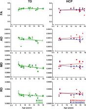
          Methods: Neuroimaging data were obtained from 36 children with HCP for both the more affected (MA) and less affected (LA) hemispheres, and, for purposes of direct comparison, between groups, 15 typically developing (TD) children. With diffusion tensor imaging (DTI), we estimated the mean fractional anisotropy (FA), axial diffusivity (AD), mean diffusivity (MD), and radial diffusivity (RD) of the corticospinal tract, parameters indicative of factors including myelination and axon density. Transcranial magnetic stimulation (TMS) was performed as a neurophysiologic measure of corticospinal tract integrity and organization. Resting motor threshold (rMT) was obtained per hemisphere, per patient.

          Results: We observed a significant AD and MD developmental trajectory, both of which were inversely related to age (decrease in AD and diffusivity corresponding to increased age) in both hemispheres of TD children ( p < 0.001). This maturation process was absent in both MA and LA hemispheres of children with HCP. Additionally, the TMS-derived previously established rMT developmental trajectory was preserved in the LA hemisphere of children with HCP ( n = 26; p < 0.0001) but this trajectory was absent in the MA hemisphere.

          Conclusions: Corticospinal tract maturation arrests in both hemispheres of children with HCP, possibly reflecting perinatal disruption of corticospinal tract myelination and axonal integrity.

          Related collections

          Most cited references23

          • Record: found
          • Abstract: found
          • Article: not found

          A report: the definition and classification of cerebral palsy April 2006.

          For a variety of reasons, the definition and the classification of cerebral palsy (CP) need to be reconsidered. Modern brain imaging techniques have shed new light on the nature of the underlying brain injury and studies on the neurobiology of and pathology associated with brain development have further explored etiologic mechanisms. It is now recognized that assessing the extent of activity restriction is part of CP evaluation and that people without activity restriction should not be included in the CP rubric. Also, previous definitions have not given sufficient prominence to the non-motor neurodevelopmental disabilities of performance and behaviour that commonly accompany CP, nor to the progression of musculoskeletal difficulties that often occurs with advancing age. In order to explore this information, pertinent material was reviewed on July 11-13, 2004 at an international workshop in Bethesda, MD (USA) organized by an Executive Committee and participated in by selected leaders in the preclinical and clinical sciences. At the workshop, it was agreed that the concept 'cerebral palsy' should be retained. Suggestions were made about the content of a revised definition and classification of CP that would meet the needs of clinicians, investigators, health officials, families and the public and would provide a common language for improved communication. Panels organized by the Executive Committee used this information and additional comments from the international community to generate a report on the Definition and Classification of Cerebral Palsy, April 2006. The Executive Committee presents this report with the intent of providing a common conceptualization of CP for use by a broad international audience.
            Bookmark
            • Record: found
            • Abstract: found
            • Article: not found

            Demyelination increases radial diffusivity in corpus callosum of mouse brain.

            Myelin damage, as seen in multiple sclerosis (MS) and other demyelinating diseases, impairs axonal conduction and can also be associated with axonal degeneration. Accurate assessments of these conditions may be highly beneficial in evaluating and selecting therapeutic strategies for patient management. Recently, an analytical approach examining diffusion tensor imaging (DTI) derived parameters has been proposed to assess the extent of axonal damage, demyelination, or both. The current study uses the well-characterized cuprizone model of experimental demyelination and remyelination of corpus callosum in mouse brain to evaluate the ability of DTI parameters to detect the progression of myelin degeneration and regeneration. Our results demonstrate that the extent of increased radial diffusivity reflects the severity of demyelination in corpus callosum of mouse brain affected by cuprizone treatment. Subsequently, radial diffusivity decreases with the progression of remyelination. Furthermore, radial diffusivity changes were specific to the time course of changes in myelin integrity as distinct from axonal injury, which was detected by betaAPP immunostaining and shown to be most extensive prior to demyelination. Radial diffusivity offers a specific assessment of demyelination and remyelination, as distinct from acute axonal damage.
              Bookmark
              • Record: found
              • Abstract: found
              • Article: not found

              Longitudinal development of human brain wiring continues from childhood into adulthood.

              Healthy human brain development is a complex process that continues during childhood and adolescence, as demonstrated by many cross-sectional and several longitudinal studies. However, whether these changes end in adolescence is not clear. We examined longitudinal white matter maturation using diffusion tensor tractography in 103 healthy subjects aged 5-32 years; each volunteer was scanned at least twice, with 221 total scans. Fractional anisotropy (FA) and mean diffusivity (MD), parameters indicative of factors including myelination and axon density, were assessed in 10 major white matter tracts. All tracts showed significant nonlinear development trajectories for FA and MD. Significant within-subject changes occurred in the vast majority of children and early adolescents, and these changes were mostly complete by late adolescence for projection and commissural tracts. However, association tracts demonstrated postadolescent within-subject maturation of both FA and MD. Diffusion parameter changes were due primarily to decreasing perpendicular diffusivity, although increasing parallel diffusivity contributed to the prolonged increases of FA in association tracts. Volume increased significantly with age for most tracts, and longitudinal measures also demonstrated postadolescent volume increases in several association tracts. As volume increases were not directly associated with either elevated FA or reduced MD between scans, the observed diffusion parameter changes likely reflect microstructural maturation of brain white matter tracts rather than just gross anatomy.
                Bookmark

                Author and article information

                Contributors
                Journal
                Front Hum Neurosci
                Front Hum Neurosci
                Front. Hum. Neurosci.
                Frontiers in Human Neuroscience
                Frontiers Media S.A.
                1662-5161
                24 July 2019
                2019
                : 13
                : 254
                Affiliations
                [1] 1Laboratory of Children’s Brain Dynamics, Division of Newborn Medicine, Boston Children’s Hospital, Harvard Medical School , Boston, MA, United States
                [2] 2Fetal-Neonatal Neuroimaging and Developmental Science Center, Division of Newborn Medicine, Boston Children’s Hospital, Harvard Medical School , Boston, MA, United States
                [3] 3Neuromodulation Program, Division of Epilepsy and Clinical Neurophysiology, Department of Neurology, Boston Children’s Hospital , Boston, MA, United States
                [4] 4F.M. Kirby Neurobiology Center, Boston Children’s Hospital , Boston, MA, United States
                [5] 5Department of Orthopedic Surgery, Boston Children’s Hospital, Harvard Medical School , Boston, MA, United States
                [6] 6Department of Radiology, Boston Children’s Hospital, Harvard Medical School , Boston, MA, United States
                [7] 7Department of Neurology, Berenson-Allen Center for Noninvasive Brain Stimulation, Division of Cognitive Neurology, Harvard Medical School and Beth Israel Deaconess Medical Center , Boston, MA, United States
                Author notes

                Edited by: Hubert Preissl, Institute for Diabetes Research and Metabolic Diseases (IDM), Germany

                Reviewed by: Giovanni Pellegrino, Montreal Neurological Institute and Hospital, McGill University, Canada; Martin Victor Sale, University of Queensland, Australia

                These authors share first authorship

                Article
                10.3389/fnhum.2019.00254
                6668599
                40986a14-6076-42e0-a849-f9deecfcb06a
                Copyright © 2019 Papadelis, Kaye, Shore, Snyder, Grant and Rotenberg.

                This is an open-access article distributed under the terms of the Creative Commons Attribution License (CC BY). The use, distribution or reproduction in other forums is permitted, provided the original author(s) and the copyright owner(s) are credited and that the original publication in this journal is cited, in accordance with accepted academic practice. No use, distribution or reproduction is permitted which does not comply with these terms.

                History
                : 27 February 2019
                : 08 July 2019
                Page count
                Figures: 4, Tables: 2, Equations: 0, References: 30, Pages: 9, Words: 5971
                Funding
                Funded by: Eunice Kennedy Shriver National Institute of Child Health and Human Development 10.13039/100009633
                Categories
                Neuroscience
                Original Research

                Neurosciences
                hemiplegic cerebral palsy,corticospinal tracts,development,maturation,transcranial magnetic stimulation

                Comments

                Comment on this article