2
views
0
recommends
+1 Recommend
0 collections
    0
    shares
      • Record: found
      • Abstract: found
      • Article: found
      Is Open Access

      Spontaneous Formation of 3D Breast Cancer Tissues on Electrospun Chitosan/Poly(ethylene oxide) Nanofibrous Scaffolds

      research-article

      Read this article at

      Bookmark
          There is no author summary for this article yet. Authors can add summaries to their articles on ScienceOpen to make them more accessible to a non-specialist audience.

          Abstract

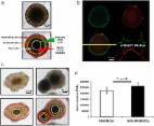
          Three-dimensional (3D) tissue culture has attracted a great deal of attention as a result of the need to replace the conventional two-dimensional cell cultures with more meaningful methods, especially for understanding the sophisticated nature of native tumor microenvironments. However, most techniques for 3D tissue culture are laborious, expensive, and limited to spheroid formation. In this study, a low-cost and highly effective nanofibrous scaffold is presented for spontaneous formation of reproducible 3D breast cancer microtissues. Experimentally, aligned and non-aligned chitosan/poly(ethylene oxide) nanofibrous scaffolds were prepared at one of two chitosan concentrations (2 and 4 wt %) and various electrospinning parameters. The resulting fabricated scaffolds (C2P1 and C4P1) were structurally and morphologically characterized, as well as analyzed in silico. The obtained data suggest that the fiber diameter, surface roughness, and scaffold wettability are tunable and can be influenced based on the chitosan concentration, electrospinning conditions, and alignment mode. To test the usefulness of the fabricated scaffolds for 3D cell culture, a breast cancer cell line (MCF-7) was cultured on their surfaces and evaluated morphologically and biochemically. The obtained data showed a higher proliferation rate for cells grown on scaffolds compared to cells grown on two-dimensional adherent plates (tissue culture plate). The MTT assay revealed that the rate of cell proliferation on nanofibrous scaffolds is statistically significantly higher compared to tissue culture plate ( P ≤ 0.001) after 14 days of culture. The formation of spheroids within the first few days of culture shows that the scaffolds effectively support 3D tissue culture from the outset of the experiment. Furthermore, 3D breast cancer tissues were spontaneously formed within 10 days of culture on aligned and non-aligned nanofibrous scaffolds, which suggests that the scaffolds imitate the in vivo extracellular matrix in the tumor microenvironment. Detailed mechanisms for the spontaneous formation of the 3D microtissues have been proposed. Our results suggest that scaffold surface topography significantly influences tissue formation and behavior of the cells.

          Related collections

          Most cited references95

          • Record: found
          • Abstract: found
          • Article: found
          Is Open Access

          Three-Dimensional in Vitro Cell Culture Models in Drug Discovery and Drug Repositioning

          Drug development is a lengthy and costly process that proceeds through several stages from target identification to lead discovery and optimization, preclinical validation and clinical trials culminating in approval for clinical use. An important step in this process is high-throughput screening (HTS) of small compound libraries for lead identification. Currently, the majority of cell-based HTS is being carried out on cultured cells propagated in two-dimensions (2D) on plastic surfaces optimized for tissue culture. At the same time, compelling evidence suggests that cells cultured in these non-physiological conditions are not representative of cells residing in the complex microenvironment of a tissue. This discrepancy is thought to be a significant contributor to the high failure rate in drug discovery, where only a low percentage of drugs investigated ever make it through the gamut of testing and approval to the market. Thus, three-dimensional (3D) cell culture technologies that more closely resemble in vivo cell environments are now being pursued with intensity as they are expected to accommodate better precision in drug discovery. Here we will review common approaches to 3D culture, discuss the significance of 3D cultures in drug resistance and drug repositioning and address some of the challenges of applying 3D cell cultures to high-throughput drug discovery.
            Bookmark
            • Record: found
            • Abstract: found
            • Article: found
            Is Open Access

            3D tumor spheroid models for in vitro therapeutic screening: a systematic approach to enhance the biological relevance of data obtained

            The potential of a spheroid tumor model composed of cells in different proliferative and metabolic states for the development of new anticancer strategies has been amply demonstrated. However, there is little or no information in the literature on the problems of reproducibility of data originating from experiments using 3D models. Our analyses, carried out using a novel open source software capable of performing an automatic image analysis of 3D tumor colonies, showed that a number of morphology parameters affect the response of large spheroids to treatment. In particular, we found that both spheroid volume and shape may be a source of variability. We also compared some commercially available viability assays specifically designed for 3D models. In conclusion, our data indicate the need for a pre-selection of tumor spheroids of homogeneous volume and shape to reduce data variability to a minimum before use in a cytotoxicity test. In addition, we identified and validated a cytotoxicity test capable of providing meaningful data on the damage induced in large tumor spheroids of up to diameter in 650 μm by different kinds of treatments.
              Bookmark
              • Record: found
              • Abstract: found
              • Article: found
              Is Open Access

              2D and 3D cell cultures – a comparison of different types of cancer cell cultures

              Cell culture is a widely used in vitro tool for improving our understanding of cell biology, tissue morphology, and mechanisms of diseases, drug action, protein production and the development of tissue engineering. Most research regarding cancer biology is based on experiments using two-dimensional (2D) cell cultures in vitro. However, 2D cultures have many limitations, such as the disturbance of interactions between the cellular and extracellular environments, changes in cell morphology, polarity, and method of division. These disadvantages led to the creation of models which are more closely able to mimic conditions in vivo. One such method is three-dimensional culture (3D). Optimisation of the culture conditions may allow for a better understanding of cancer biology and facilitate the study of biomarkers and targeting therapies. In this review, we compare 2D and 3D cultures in vitro as well as different versions of 3D cultures.
                Bookmark

                Author and article information

                Journal
                ACS Omega
                ACS Omega
                ao
                acsodf
                ACS Omega
                American Chemical Society
                2470-1343
                05 January 2022
                18 January 2022
                : 7
                : 2
                : 2114-2126
                Affiliations
                []Environmental and Smart Technology Group (ESTG), Faculty of Science, Fayoum University , 63514 Fayoum, Egypt
                []Chemistry Department, Faculty of Science, Helwan University , Ain Helwan, 11795 Cairo, Egypt
                [§ ]Department of Applied Biochemistry, Institute of Biotechnology, Technische Universität Berlin , 13355 Berlin, Germany
                []Nanotechnology Research Center (NTRC), The British University in Egypt (BUE) , El-Sherouk City, 11837 Cairo, Egypt
                []Department of Medicinal Drugs, National Research Center , 12622 Giza, Egypt
                [# ]Materials Science & Engineering Department, School of Innovative Design Engineering, Egypt-Japan University of Science and Technology (E-JUST) , 21934 Alexandria, Egypt
                Author notes
                Author information
                https://orcid.org/0000-0002-4859-5264
                https://orcid.org/0000-0002-1469-0052
                https://orcid.org/0000-0003-1825-6752
                Article
                10.1021/acsomega.1c05646
                8771982
                35071900
                40dd2d79-81a7-4779-a4d4-b7fd0ab9b3f7
                © 2022 The Authors. Published by American Chemical Society

                Permits non-commercial access and re-use, provided that author attribution and integrity are maintained; but does not permit creation of adaptations or other derivative works ( https://creativecommons.org/licenses/by-nc-nd/4.0/).

                History
                : 09 October 2021
                : 22 December 2021
                Funding
                Funded by: Arab-German Young Academy of Sciences and Humanities, doi NA;
                Award ID: NA
                Categories
                Article
                Custom metadata
                ao1c05646
                ao1c05646

                Comments

                Comment on this article