2
views
0
recommends
+1 Recommend
0 collections
    0
    shares
      • Record: found
      • Abstract: found
      • Article: found
      Is Open Access

      Involvement of the Nucleus Incertus and Relaxin-3/RXFP3 Signaling System in Explicit and Implicit Memory

      research-article

      Read this article at

      Bookmark
          There is no author summary for this article yet. Authors can add summaries to their articles on ScienceOpen to make them more accessible to a non-specialist audience.

          Abstract

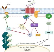
          Telencephalic cognitive and emotional circuits/functions are strongly modulated by subcortical inputs. The main focus of past research on the nature of this modulation has been on the widespread monoamine projections to the telencephalon. However, the nucleus incertus (NI) of the pontine tegmentum provides a strong GABAergic and peptidergic innervation of the hippocampus, basal forebrain, amygdala, prefrontal cortex, and related regions; and represents a parallel source of ascending modulation of cognitive and emotional domains. NI GABAergic neurons express multiple peptides, including neuromedin-B, cholecystokinin, and relaxin-3, and receptors for stress and arousal transmitters, including corticotrophin-releasing factor and orexins/hypocretins. A functional relationship exists between NI neurons and their associated peptides, relaxin-3 and neuromedin-B, and hippocampal theta rhythm, which in turn, has a key role in the acquisition and extinction of declarative and emotional memories. Furthermore, RXFP3, the cognate receptor for relaxin-3, is a G i/o protein-coupled receptor, and its activation inhibits the cellular accumulation of cAMP and induces phosphorylation of ERK, processes associated with memory formation in the hippocampus and amygdala. Therefore, this review summarizes the role of NI transmitter systems in relaying stress- and arousal-related signals to the higher neural circuits and processes associated with memory formation and retrieval.

          Related collections

          Most cited references133

          • Record: found
          • Abstract: found
          • Article: not found

          Memory, navigation and theta rhythm in the hippocampal-entorhinal system.

          Theories on the functions of the hippocampal system are based largely on two fundamental discoveries: the amnestic consequences of removing the hippocampus and associated structures in the famous patient H.M. and the observation that spiking activity of hippocampal neurons is associated with the spatial position of the rat. In the footsteps of these discoveries, many attempts were made to reconcile these seemingly disparate functions. Here we propose that mechanisms of memory and planning have evolved from mechanisms of navigation in the physical world and hypothesize that the neuronal algorithms underlying navigation in real and mental space are fundamentally the same. We review experimental data in support of this hypothesis and discuss how specific firing patterns and oscillatory dynamics in the entorhinal cortex and hippocampus can support both navigation and memory.
            Bookmark
            • Record: found
            • Abstract: not found
            • Article: not found

            The hippocampus as a spatial map. Preliminary evidence from unit activity in the freely-moving rat

              Bookmark
              • Record: found
              • Abstract: found
              • Article: not found

              Dissociable roles of prelimbic and infralimbic cortices, ventral hippocampus, and basolateral amygdala in the expression and extinction of conditioned fear.

              Current models of conditioned fear expression and extinction involve the basolateral amygdala (BLA), ventral medial prefrontal cortex (vmPFC), and the hippocampus (HPC). There is some disagreement with respect to the specific roles of these structures, perhaps due to subregional differences within each area. For example, growing evidence suggests that infralimbic (IL) and prelimbic (PL) subregions of vmPFC have opposite influences on fear expression. Moreover, it is the ventral HPC (vHPC), rather than the dorsal HPC, that projects to vmPFC and BLA. To help determine regional specificity, we used small doses of the GABA(A) agonist muscimol to selectively inactivate IL, PL, BLA, or vHPC in an auditory fear conditioning and extinction paradigm. Infusions were performed prior to extinction training, allowing us to assess the effects on both fear expression and subsequent extinction memory. Inactivation of IL had no effect on fear expression, but impaired the within-session acquisition of extinction as well as extinction memory. In contrast, inactivation of PL impaired fear expression, but had no effect on extinction memory. Inactivation of the BLA or vHPC impaired both fear expression and extinction memory. Post-extinction inactivations had no effect in any structure. We suggest a model in which amygdala-dependent fear expression is modulated by inputs from PL and vHPC, whereas extinction memory requires extinction-induced plasticity in IL, BLA, and/or vHPC.
                Bookmark

                Author and article information

                Contributors
                Journal
                Front Neuroanat
                Front Neuroanat
                Front. Neuroanat
                Frontiers in Neuroanatomy
                Frontiers Media S.A.
                1662-5129
                18 March 2021
                2021
                : 15
                : 637922
                Affiliations
                [1] 1Unitat Predepartamental de Medicina, Facultat de Ciències de la Salut, Universitat Jaume I , Castelló de la Plana, Spain
                [2] 2Centro de Investigación Biomédica en Red de Salud Mental, CIBERSAM , Madrid, Spain
                [3] 3Department of Clinical Neurosciences, UK Dementia Research Institute, University of Cambridge , Cambridge, United Kingdom
                [4] 4The Florey Institute for Neuroscience and Mental Health, The University of Melbourne , Parkville, VIC, Australia
                Author notes

                Edited by: Alexander C. Jackson, University of Connecticut, United States

                Reviewed by: Eric E. Turner, Seattle Children’s Hospital, United States; Christopher S. Leonard, New York Medical College, United States

                *Correspondence: Francisco E. Olucha-Bordonau folucha@ 123456uji.es
                Article
                10.3389/fnana.2021.637922
                8044989
                33867946
                411b41db-738b-4d2c-83b9-555afe9f3295
                Copyright © 2021 Gil-Miravet, Mañas-Ojeda, Ros-Bernal, Castillo-Gómez, Albert-Gascó, Gundlach and Olucha-Bordonau.

                This is an open-access article distributed under the terms of the Creative Commons Attribution License (CC BY). The use, distribution or reproduction in other forums is permitted, provided the original author(s) and the copyright owner(s) are credited and that the original publication in this journal is cited, in accordance with accepted academic practice. No use, distribution or reproduction is permitted which does not comply with these terms.

                History
                : 04 December 2020
                : 26 February 2021
                Page count
                Figures: 4, Tables: 0, Equations: 0, References: 133, Pages: 15, Words: 11961
                Categories
                Neuroscience
                Review

                Neurosciences
                neuropeptide,rxfp3,gaba,brainstem,amygdala,hippocampus
                Neurosciences
                neuropeptide, rxfp3, gaba, brainstem, amygdala, hippocampus

                Comments

                Comment on this article