18
views
0
recommends
+1 Recommend
0 collections
    0
    shares
      • Record: found
      • Abstract: found
      • Article: found
      Is Open Access

      LATS2 overexpression attenuates the therapeutic resistance of liver cancer HepG2 cells to sorafenib-mediated death via inhibiting the AMPK–Mfn2 signaling pathway

      research-article
      1 , 2 , 3 , 1 ,
      Cancer Cell International
      BioMed Central
      HCC, Mitophagy, Sorafenib, AMPK pathway, LATS2

      Read this article at

      Bookmark
          There is no author summary for this article yet. Authors can add summaries to their articles on ScienceOpen to make them more accessible to a non-specialist audience.

          Abstract

          Background

          Effective therapy for hepatocellular carcinoma (HCC) is currently an imperative issue, and sorafenib is a first-line drug for the treatment of HCC. However, the clinical benefit of sorafenib is often impaired by drug resistance. Accordingly, the present study was conducted to investigate the molecular mechanisms involving sorafenib resistance, with a focus on large tumor suppressor 2 (LATS2) and mitophagy.

          Methods

          HepG2 liver cancer cells were treated with sorafenib and infected with adenovirus-loaded LATS2 (Ad-LATS2). Cell death, proliferation and migration were measured via western blotting analysis, immunofluorescence and qPCR. Mitochondrial function and mitophagy were determined via western blotting and immunofluorescence.

          Results

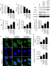
          Our data indicated that LATS2 expression was repressed by sorafenib treatment, and overexpression of LATS2 could further enhance sorafenib-mediated apoptosis in HepG2 liver cancer cells. At the molecular level, mitochondrial stress was triggered by sorafenib treatment, as evidenced by decreased mitochondrial membrane potential, increased mitochondrial ROS production, more cyc-c release into the nucleus, and elevated mitochondrial pro-apoptotic proteins. However, in response to mitochondrial damage, mitophagy was activated by sorafenib treatment, whereas LATS2 overexpression effectively inhibited mitophagy activity and thus augmented sorafenib-mediated mitochondrial stress. Subsequently, we also demonstrated that the AMPK–MFN2 signaling pathway was involved in mitophagy regulation after exposure to sorafenib treatment and/or LATS2 overexpression. Inhibition of the AMPK pathway interrupted mitophagy and thus enhanced the antitumor property of sorafenib, similar to the results obtained via overexpression of LATS2.

          Conclusions

          Altogether, our findings revealed the importance of the LATS2/AMPK/MFN2/mitophagy axis in understanding sorafenib resistance mechanisms, with a potential application to increase the sensitivity response of sorafenib in the treatment of liver cancer.

          Related collections

          Most cited references59

          • Record: found
          • Abstract: found
          • Article: found
          Is Open Access

          Pathogenesis of cardiac ischemia reperfusion injury is associated with CK2α-disturbed mitochondrial homeostasis via suppression of FUNDC1-related mitophagy

          Disturbed mitochondrial homeostasis contributes to the pathogenesis of cardiac ischemia reperfusion (IR) injury, although the underlying mechanism remains elusive. Here, we demonstrated that casein kinase 2α (CK2α) was upregulated following acute cardiac IR injury. Increased CK2α was shown to be instrumental to mitochondrial damage, cardiomyocyte death, infarction area expansion and cardiac dysfunction, whereas cardiac-specific CK2α knockout (CK2α CKO ) mice were protected against IR injury and mitochondrial damage. Functional assay indicated that CK2α enhanced the phosphorylation (inactivation) of FUN14 domain containing 1 (FUNDC1) via post-transcriptional modification at Ser13, thus effectively inhibiting mitophagy. Defective mitophagy failed to remove damaged mitochondria induced by IR injury, resulting in mitochondrial genome collapse, electron transport chain complex (ETC) inhibition, mitochondrial biogenesis arrest, cardiolipin oxidation, oxidative stress, mPTP opening, mitochondrial debris accumulation and eventually mitochondrial apoptosis. In contrast, loss of CK2α reversed the FUNDC1-mediated mitophagy, providing a survival advantage to myocardial tissue following IR stress. Interestingly, mice deficient in both CK2α and FUNDC1 failed to show protection against IR injury and mitochondrial damage through a mechanism possible attributed to lack of mitophagy. Taken together, our results confirmed that CK2α serves as a negative regulator of mitochondrial homeostasis via suppression of FUNDC1-required mitophagy, favoring the development of cardiac IR injury.
            Bookmark
            • Record: found
            • Abstract: found
            • Article: found
            Is Open Access

            Melatonin protects cardiac microvasculature against ischemia/reperfusion injury via suppression of mitochondrial fission‐VDAC1‐HK2‐mPTP‐mitophagy axis

            Abstract The cardiac microvascular system, which is primarily composed of monolayer endothelial cells, is the site of blood supply and nutrient exchange to cardiomyocytes. However, microvascular ischemia/reperfusion injury (IRI) following percutaneous coronary intervention is a woefully neglected topic, and few strategies are available to reverse such pathologies. Here, we studied the effects of melatonin on microcirculation IRI and elucidated the underlying mechanism. Melatonin markedly reduced infarcted area, improved cardiac function, restored blood flow, and lower microcirculation perfusion defects. Histological analysis showed that cardiac microcirculation endothelial cells (CMEC) in melatonin‐treated mice had an unbroken endothelial barrier, increased endothelial nitric oxide synthase expression, unobstructed lumen, reduced inflammatory cell infiltration, and less endothelial damage. In contrast, AMP‐activated protein kinase α (AMPKα) deficiency abolished the beneficial effects of melatonin on microvasculature. In vitro, IRI activated dynamin‐related protein 1 (Drp1)‐dependent mitochondrial fission, which subsequently induced voltage‐dependent anion channel 1 (VDAC1) oligomerization, hexokinase 2 (HK2) liberation, mitochondrial permeability transition pore (mPTP) opening, PINK1/Parkin upregulation, and ultimately mitophagy‐mediated CMEC death. However, melatonin strengthened CMEC survival via activation of AMPKα, followed by p‐Drp1S616 downregulation and p‐Drp1S37 upregulation, which blunted Drp1‐dependent mitochondrial fission. Suppression of mitochondrial fission by melatonin recovered VDAC1‐HK2 interaction that prevented mPTP opening and PINK1/Parkin activation, eventually blocking mitophagy‐mediated cellular death. In summary, this study confirmed that melatonin protects cardiac microvasculature against IRI. The underlying mechanism may be attributed to the inhibitory effects of melatonin on mitochondrial fission‐VDAC1‐HK2‐mPTP‐mitophagy axis via activation of AMPKα.
              Bookmark
              • Record: found
              • Abstract: found
              • Article: not found

              NR4A1 aggravates the cardiac microvascular ischemia reperfusion injury through suppressing FUNDC1-mediated mitophagy and promoting Mff-required mitochondrial fission by CK2α.

              Mitochondrial fission and mitophagy are considered key processes involved in the pathogenesis of cardiac microvascular ischemia reperfusion (IR) injury although the upstream regulatory mechanism for fission and mitophagy still remains unclear. Herein, we reported that NR4A1 was significantly upregulated following cardiac microvascular IR injury, and its level was positively correlated with microvascular collapse, endothelial cellular apoptosis and mitochondrial damage. However, NR4A1-knockout mice exhibited resistance against the acute microvascular injury and mitochondrial dysfunction compared with the wild-type mice. Functional studies illustrated that IR injury increased NR4A1 expression, which activated serine/threonine kinase casein kinase2 α (CK2α). CK2α promoted phosphorylation of mitochondrial fission factor (Mff) and FUN14 domain-containing 1 (FUNDC1). Phosphorylated activation of Mff enhanced the cytoplasmic translocation of Drp1 to the mitochondria, leading to fatal mitochondrial fission. Excessive fission disrupted mitochondrial function and structure, ultimately triggering mitochondrial apoptosis. In addition, phosphorylated inactivation of FUNDC1 failed to launch the protective mitophagy process, resulting in the accumulation of damaged mitochondria and endothelial apoptosis. By facilitating Mff-mediated mitochondrial fission and FUNDC1-required mitophagy, NR4A1 disturbed mitochondrial homeostasis, enhanced endothelial apoptosis and provoked microvascular dysfunction. In summary, our data illustrated that NR4A1 serves as a novel culprit factor in cardiac microvascular IR injury that operates through synchronous elevation of fission and suppression of mitophagy. Novel therapeutic strategies targeting the balance among NR4A1, fission and mitophagy might provide survival advantage to microvasculature following IR stress.
                Bookmark

                Author and article information

                Contributors
                16697974@qq.com
                Journal
                Cancer Cell Int
                Cancer Cell Int
                Cancer Cell International
                BioMed Central (London )
                1475-2867
                18 March 2019
                18 March 2019
                2019
                : 19
                : 60
                Affiliations
                [1 ]GRID grid.452829.0, Department of Hepatopancreatobiliary Medicine, , The Second Hospital of Jilin University, ; Changchun, 130000 China
                [2 ]GRID grid.452829.0, Department of Pharmacy, , The Second Hospital of Jilin University, ; Changchun, 130000 China
                [3 ]GRID grid.452829.0, Department of Anesthesiology, , The Second Hospital of Jilin University, ; Changchun, 130000 China
                Article
                778
                10.1186/s12935-019-0778-1
                6423758
                30923462
                41c28e3d-3a4c-4262-b578-a53e1afebb32
                © The Author(s) 2019

                Open AccessThis article is distributed under the terms of the Creative Commons Attribution 4.0 International License ( http://creativecommons.org/licenses/by/4.0/), which permits unrestricted use, distribution, and reproduction in any medium, provided you give appropriate credit to the original author(s) and the source, provide a link to the Creative Commons license, and indicate if changes were made. The Creative Commons Public Domain Dedication waiver ( http://creativecommons.org/publicdomain/zero/1.0/) applies to the data made available in this article, unless otherwise stated.

                History
                : 8 February 2019
                : 8 March 2019
                Categories
                Primary Research
                Custom metadata
                © The Author(s) 2019

                Oncology & Radiotherapy
                hcc,mitophagy,sorafenib,ampk pathway,lats2
                Oncology & Radiotherapy
                hcc, mitophagy, sorafenib, ampk pathway, lats2

                Comments

                Comment on this article