13
views
0
recommends
+1 Recommend
0 collections
    0
    shares
      • Record: found
      • Abstract: found
      • Article: found
      Is Open Access

      PAR1 Scaffolds TGFβRII to Downregulate TGF-β Signaling and Activate ESC Differentiation to Endothelial Cells

      research-article

      Read this article at

      Bookmark
          There is no author summary for this article yet. Authors can add summaries to their articles on ScienceOpen to make them more accessible to a non-specialist audience.

          Summary

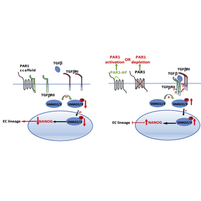
          We studied the function of the G-protein-coupled receptor PAR1 in mediating the differentiation of mouse embryonic stem cells (mESCs) to endothelial cells (ECs) that are capable of inducing neovascularization. We observed that either deletion or activation of PAR1 suppressed mouse embryonic stem cell (mESC) differentiation to ECs and neovascularization in mice. This was mediated by induction of TGFβRII/TGFβRI interaction, forming an active complex, which in turn induced SMAD2 phosphorylation. Inhibition of TGF-β signaling in PAR1-deficient mESCs restored the EC differentiation potential of mESCs. Thus, PAR1 in its inactive unligated state functions as a scaffold for TGFβRII to downregulate TGF-β signaling, and thereby promote ESC transition to functional ECs. The PAR1 scaffold function in ESCs is an essential mechanism for dampening TGF-β signaling and regulating ESC differentiation.

          Graphical Abstract

          Highlights

          • ESC differentiation to ECs is regulated by PAR1 activity and expression in mESCs

          • Deletion of PAR1 suppresses EC generation and neovessel formation

          • PAR1 acts as a scaffolding partner for TGFβRII and suppresses TGF-β signaling in ESCs

          Abstract

          In this article, Malik and colleagues show that the G-protein-coupled receptor PAR1 can act as a scaffold for the TGF-β receptor TGFβRII and thereby suppress downstream TGF-β signaling in embryonic stem cells undergoing endothelial differentiation. Thus, PAR1 functions as a rheostat controlling TGF-β signaling and the generation of functional endothelial cells from embryonic stem cells.

          Related collections

          Most cited references26

          • Record: found
          • Abstract: found
          • Article: not found

          Thrombin signalling and protease-activated receptors.

          S Coughlin (2000)
          How does the coagulation protease thrombin regulate cellular behaviour? The protease-activated receptors (PARs) provide one answer. In concert with the coagulation cascade, these receptors provide an elegant mechanism linking mechanical information in the form of tissue injury or vascular leakage to cellular responses. Roles for PARs are beginning to emerge in haemostasis and thrombosis, inflammation, and perhaps even blood vessel development.
            Bookmark
            • Record: found
            • Abstract: found
            • Article: not found

            TGFbeta/activin/nodal signaling is necessary for the maintenance of pluripotency in human embryonic stem cells.

            Human embryonic stem cells (hESCs) self-renew indefinitely and give rise to derivatives of all three primary germ layers, yet little is known about the signaling cascades that govern their pluripotent character. Because it plays a prominent role in the early cell fate decisions of embryonic development, we have examined the role of TGFbeta superfamily signaling in hESCs. We found that, in undifferentiated cells, the TGFbeta/activin/nodal branch is activated (through the signal transducer SMAD2/3) while the BMP/GDF branch (SMAD1/5) is only active in isolated mitotic cells. Upon early differentiation, SMAD2/3 signaling is decreased while SMAD1/5 signaling is activated. We next tested the functional role of TGFbeta/activin/nodal signaling in hESCs and found that it is required for the maintenance of markers of the undifferentiated state. We extend these findings to show that SMAD2/3 activation is required downstream of WNT signaling, which we have previously shown to be sufficient to maintain the undifferentiated state of hESCs. Strikingly, we show that in ex vivo mouse blastocyst cultures, SMAD2/3 signaling is also required to maintain the inner cell mass (from which stem cells are derived). These data reveal a crucial role for TGFbeta signaling in the earliest stages of cell fate determination and demonstrate an interconnection between TGFbeta and WNT signaling in these contexts.
              Bookmark
              • Record: found
              • Abstract: found
              • Article: not found

              Wnt signaling in stem and cancer stem cells.

              The functional versatility of Wnt/β-catenin signaling can be seen by its ability to act in stem cells of the embryo and of the adult as well as in cancer stem cells. During embryogenesis, stem cells demonstrate a requirement for β-catenin in mediating the response to Wnt signaling for their maintenance and transition from a pluripotent state. In adult stem cells, Wnt signaling functions at various hierarchical levels to contribute to specification of different tissues. This has raised the possibility that the tightly regulated self-renewal mediated by Wnt signaling in stem and progenitor cells is subverted in cancer cells to allow malignant progression. Intensive work is currently being performed to resolve how intrinsic and extrinsic factors that regulate Wnt/β-catenin signaling coordinate the stem and cancer stem cell states. Copyright © 2013 Elsevier Ltd. All rights reserved.
                Bookmark

                Author and article information

                Contributors
                Journal
                Stem Cell Reports
                Stem Cell Reports
                Stem Cell Reports
                Elsevier
                2213-6711
                17 November 2016
                13 December 2016
                17 November 2016
                : 7
                : 6
                : 1050-1058
                Affiliations
                [1 ]Department of Pharmacology, University of Illinois, College of Medicine, 835 South Wolcott Avenue, Room E403, Chicago, IL 60612, USA
                [2 ]The Center for Lung and Vascular Biology, University of Illinois, College of Medicine, 835 South Wolcott Avenue, Room E403, Chicago, IL 60612, USA
                [3 ]Department of Pharmacology, Max-Planck-Institute for Heart and Lung Research, Bad Nauheim 61231, Germany
                [4 ]Feinberg School of Medicine, Northwestern University, Chicago, IL 60611, USA
                Author notes
                []Corresponding author abmalik@ 123456uic.edu
                Article
                S2213-6711(16)30244-2
                10.1016/j.stemcr.2016.10.006
                5161529
                27866874
                4276aee0-e833-4784-845a-64add267b2b4
                © 2016 The Author(s)

                This is an open access article under the CC BY license (http://creativecommons.org/licenses/by/4.0/).

                History
                : 5 January 2016
                : 14 October 2016
                : 17 October 2016
                Categories
                Article

                gpcr,g-protein-coupled receptor,stem cells,embryonic stem cells,endothelial cells,cell differentiation,par1,tgf-β signaling,scaffolding protein

                Comments

                Comment on this article