14
views
0
recommends
+1 Recommend
0 collections
    3
    shares
      • Record: found
      • Abstract: found
      • Article: found
      Is Open Access

      Component corneal surgery: An update

      review-article

      Read this article at

      Bookmark
          There is no author summary for this article yet. Authors can add summaries to their articles on ScienceOpen to make them more accessible to a non-specialist audience.

          Abstract

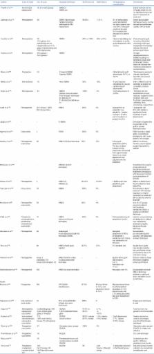
          Several decades ago, penetrating keratoplasty was a challenge to corneal surgeons. Constant effort by the corneal surgeon to improve the outcomes as well as utilization of the available resources has led to a revolutionary change in the field of keratoplasty. All these efforts have led to the evolution of techniques that allow a corneal surgeon to disease-specific transplant of individual layers of corneal “so-called component corneal surgery” depending on the layer of cornea affected. This has led to an improvement in corneal graft survival as well as a better utilization of corneal tissues. This article reviews the currently available literature on component corneal surgeries and provides an update on the available techniques.

          Related collections

          Most cited references98

          • Record: found
          • Abstract: found
          • Article: not found

          Descemet membrane endothelial keratoplasty (DMEK).

          To describe Descemet membrane endothelial keratoplasty (DMEK) with organ cultured Descemet membrane (DM) in a human cadaver eye model and a patient with Fuchs endothelial dystrophy. In 10 human cadaver eyes and 1 patient eye, a 3.5-mm clear corneal tunnel incision was made. The anterior chamber was filled with air, and the DM was stripped off from the posterior stroma. From organ-cultured donor corneo-scleral rims, 9.0-mm-diameter "DM rolls" were harvested. Each donor DM roll was inserted into a recipient anterior chamber, positioned onto the posterior stroma, and kept in position by completely filling the anterior chamber with air for 30 minutes. In all recipient eyes, the donor DM maintained its position after a 30-minute air-fill of the anterior chamber followed by an air-liquid exchange. In the patient's eye, 1 week after transplantation, best-corrected visual acuity was 1.0 (20/20) with the patient's preoperative refraction, and the endothelial cell density averaged 2350 cells/mm. DMEK may provide quick visual rehabilitation in the treatment of corneal endothelial disorders by transplantation of an organ-cultured DM transplanted through a clear corneal tunnel incision. DMEK may be a highly accessible procedure to corneal surgeons, because donor DM sheets can be prepared from preserved corneo-scleral rims.
            Bookmark
            • Record: found
            • Abstract: found
            • Article: not found

            Preliminary clinical results of Descemet membrane endothelial keratoplasty.

            To describe the preliminary clinical results of selective transplantation of organ cultured, donor Descemet membrane (DM) carrying autologous corneal endothelium through a 3.5-mm incision, tentatively named Descemet membrane endothelial keratoplasty (DMEK), for the management of corneal endothelial disorders. Nonrandomized clinical study. In 10 patients with Fuchs endothelial dystrophy or pseudophakic bullous keratopathy, DMEK was performed. A 3.5-mm clear corneal tunnel incision was made, the anterior chamber was filled with air, and DM was stripped off from the posterior stroma. A 9.0-mm diameter DM roll was harvested from an organ cultured donor corneo-scleral rim, and inserted into a recipient anterior chamber. The donor tissue was gently unfolded, positioned onto the posterior stroma, and secured by completely filling the anterior chamber with air for 30 minutes. At one month, six eyes had a best-corrected visual acuity of 0.5 (20/40) or better, and three eyes reached 1.0 (20/20). At six months, the endothelial cell density averaged 2030 (+/-373) cells/mm(2) (n = 7). Three eyes showed a complete detachment of the donor tissue in the early postoperative course that was managed by removal of the transplant and a secondary Descemet stripping endothelial keratoplasty procedure. DMEK may have potential to become the most preferable technique to manage corneal endothelial disorders, because it provides quick and nearly complete visual rehabilitation. Because the donor tissue required can be prepared from organ cultured corneo-scleral rims, the procedure may be readily accessible to most corneal surgeons.
              Bookmark
              • Record: found
              • Abstract: found
              • Article: not found

              Deep lamellar keratoplasty with complete removal of pathological stroma for vision improvement.

              Deep lamellar keratoplasty (DLK) was performed to restore visual acuity in 120 eyes with corneal stromal opacification. DLK is believed to be an effective treatment in eyes in which endothelial cell function had been preserved, and in which there was no epithelial or stromal oedema. The purpose of this study was to evaluate the effectiveness of this treatment. The stroma was excised to the extent that only Descemet's membrane remained, at least in the optical zone. Donor corneas of full, or almost full, thickness with Descemet's membrane removed, or which had been lathed to a thickness of 0.4 mm from the endothelial side, were attached by suturing. In 113 eyes which were observed for 6 months or more postoperatively in which average prospective visual acuity was 0.09, average postoperative visual acuity improved to 0.6. Specular microscopy 1 month postoperatively revealed average endothelial cell counts of 2225 (SD 659)/mm2, while 24 months postoperatively this value was 1937 (642)/mm2 (cell loss 13%). Puncturing of Descemet's membrane during surgery occurred in 47 of 120 eyes (39.2%), but after 12 months, there was no difference in visual acuity or number of endothelial cells between these eyes and those in which no puncturing had occurred. There was no postoperative endothelial rejection reaction with DLK, and restoration of postoperative visual acuity was quite adequate. Compared with penetrating keratoplasty, DLK allows endothelial cell counts to be maintained for a longer period. In addition, results can be expected to be more consistent over the long term with DLK.
                Bookmark

                Author and article information

                Journal
                Indian J Ophthalmol
                Indian J Ophthalmol
                IJO
                Indian Journal of Ophthalmology
                Medknow Publications & Media Pvt Ltd (India )
                0301-4738
                1998-3689
                August 2017
                : 65
                : 8
                : 658-672
                Affiliations
                [1]Dr. Rajendra Prasad Centre for Ophthalmic Sciences, All India Institute of Medical Sciences, New Delhi, India
                Author notes
                Correspondence to: Prof. Namrata Sharma, Cornea, Cataract and Refractive Surgery Services, Dr. Rajendra Prasad Centre for Ophthalmic Sciences, All India Institute of Medical Sciences, New Delhi, India. E-mail: namrata.sharma@ 123456gmail.com
                Article
                IJO-65-658
                10.4103/ijo.IJO_582_17
                5598175
                28820150
                44bdbebc-6c81-4ac0-aaa8-4456c32af225
                Copyright: © 2017 Indian Journal of Ophthalmology

                This is an open access article distributed under the terms of the Creative Commons Attribution-NonCommercial-ShareAlike 3.0 License, which allows others to remix, tweak, and build upon the work non-commercially, as long as the author is credited and the new creations are licensed under the identical terms.

                History
                : 07 July 2017
                : 29 July 2017
                Categories
                Review Article

                Ophthalmology & Optometry
                component corneal surgery,endothelial keratoplasty,keratoplasty,lamellar keratoplasty

                Comments

                Comment on this article