3
views
0
recommends
+1 Recommend
0 collections
    0
    shares
      • Record: found
      • Abstract: found
      • Article: found
      Is Open Access

      Development of a simplified model and nomogram in preoperative diagnosis of pediatric chronic cholangitis with pancreaticobiliary maljunction using clinical variables and MRI radiomics

      research-article

      Read this article at

      Bookmark
          There is no author summary for this article yet. Authors can add summaries to their articles on ScienceOpen to make them more accessible to a non-specialist audience.

          Abstract

          Objective

          The aim of this study was to develop a model that combines clinically relevant features with radiomics signature based on magnetic-resonance imaging (MRI) for diagnosis of chronic cholangitis in pancreaticobiliary maljunction (PBM) children.

          Methods

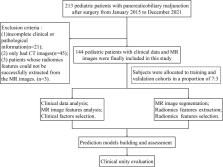
          A total of 144 subjects from two institutions confirmed PBM were included in this study. Clinical characteristics and MRI features were evaluated to build a clinical model. Radiomics features were extracted from the region of interest manually delineated on T2-weighted imaging. A radiomics signature was developed by the selected radiomics features using the least absolute shrinkage and selection operator and then a radiomics score (Rad-score) was calculated. We constructed a combined model incorporating clinical factors and Rad-score by multivariate logistic regression analysis. The combined model was visualized as a radiomics nomogram to achieve model visualization and provide clinical utility. Receiver operating curve analysis and decision curve analysis (DCA) were used to evaluate the diagnostic performance.

          Results

          Jaundice, protein plug, and ascites were selected as key clinical variables. Eight radiomics features were combined to construct the radiomics signature. The combined model showed superior predictive performance compared with the clinical model alone (AUC in the training cohort: 0.891 vs. 0.767, the validation cohort: 0.858 vs. 0.731), and the difference was significant ( p = 0.002, 0.028) in the both cohorts. DCA confirmed the clinical utility of the radiomics nomogram.

          Conclusion

          The proposed model that combines key clinical variables and radiomics signature is helpful in the diagnosis of chronic cholangitis in PBM children.

          Supplementary Information

          The online version contains supplementary material available at 10.1186/s13244-023-01383-z.

          Key points

          • Conventional imaging modalities were not powerful enough to diagnose chronic cholangitis.

          • The radiomics signature based on T2-weighted MR images performed well in diagnosing chronic cholangitis.

          • Associating the radiomics signature with clinical factors improved the diagnosis performance of chronic cholangitis.

          Supplementary Information

          The online version contains supplementary material available at 10.1186/s13244-023-01383-z.

          Related collections

          Most cited references33

          • Record: found
          • Abstract: not found
          • Article: not found

          The Measurement of Observer Agreement for Categorical Data

            Bookmark
            • Record: found
            • Abstract: found
            • Article: not found

            Radiomics: Images Are More than Pictures, They Are Data

            This report describes the process of radiomics, its challenges, and its potential power to facilitate better clinical decision making, particularly in the care of patients with cancer.
              Bookmark
              • Record: found
              • Abstract: found
              • Article: not found

              Radiomics: extracting more information from medical images using advanced feature analysis.

              Solid cancers are spatially and temporally heterogeneous. This limits the use of invasive biopsy based molecular assays but gives huge potential for medical imaging, which has the ability to capture intra-tumoural heterogeneity in a non-invasive way. During the past decades, medical imaging innovations with new hardware, new imaging agents and standardised protocols, allows the field to move towards quantitative imaging. Therefore, also the development of automated and reproducible analysis methodologies to extract more information from image-based features is a requirement. Radiomics--the high-throughput extraction of large amounts of image features from radiographic images--addresses this problem and is one of the approaches that hold great promises but need further validation in multi-centric settings and in the laboratory. Copyright © 2011 Elsevier Ltd. All rights reserved.
                Bookmark

                Author and article information

                Contributors
                gwlsuzhou@163.com
                Journal
                Insights Imaging
                Insights Imaging
                Insights into Imaging
                Springer Vienna (Vienna )
                1869-4101
                8 March 2023
                8 March 2023
                December 2023
                : 14
                : 41
                Affiliations
                [1 ]GRID grid.452253.7, ISNI 0000 0004 1804 524X, Department of Radiology, , Children’s Hospital of Soochow University, ; Suzhou, 215025 China
                [2 ]GRID grid.460138.8, Department of Radiology, , Xuzhou Children’s Hospital, ; Xuzhou, 221002 China
                [3 ]GRID grid.452253.7, ISNI 0000 0004 1804 524X, Pediatric Surgery, , Children’s Hospital of Soochow University, ; Suzhou, 215025 China
                Author information
                http://orcid.org/0000-0003-3849-2904
                Article
                1383
                10.1186/s13244-023-01383-z
                9992494
                36882647
                461442f2-a95e-418e-99d5-063196ab1323
                © The Author(s) 2023

                Open AccessThis article is licensed under a Creative Commons Attribution 4.0 International License, which permits use, sharing, adaptation, distribution and reproduction in any medium or format, as long as you give appropriate credit to the original author(s) and the source, provide a link to the Creative Commons licence, and indicate if changes were made. The images or other third party material in this article are included in the article's Creative Commons licence, unless indicated otherwise in a credit line to the material. If material is not included in the article's Creative Commons licence and your intended use is not permitted by statutory regulation or exceeds the permitted use, you will need to obtain permission directly from the copyright holder. To view a copy of this licence, visit http://creativecommons.org/licenses/by/4.0/.

                History
                : 28 September 2022
                : 4 February 2023
                Funding
                Funded by: the National Natural Science Foundation of China
                Award ID: 81971685
                Award Recipient :
                Funded by: Scientific research project of Jiangsu Provincial Health Commission
                Award ID: ZD2022015
                Award Recipient :
                Categories
                Original Article
                Custom metadata
                © The Author(s) 2023

                Radiology & Imaging
                pancreaticobiliary maljunction,children,magnetic resonance imaging,radiomics,nomogram

                Comments

                Comment on this article