19
views
0
recommends
+1 Recommend
0 collections
    0
    shares
      • Record: found
      • Abstract: found
      • Article: found
      Is Open Access

      Aggravation of collagen-induced arthritis by orally administered Porphyromonas gingivalis through modulation of the gut microbiota and gut immune system

      research-article

      Read this article at

      Bookmark
          There is no author summary for this article yet. Authors can add summaries to their articles on ScienceOpen to make them more accessible to a non-specialist audience.

          Abstract

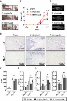
          Porhyromonas gingivalis, a causative bacterium of periodontitis, is implicated in the etiology of rheumatoid arthritis (RA), mainly because of expressing peptidyl arginine deiminase (PAD) that generates RA-related autoantigens. However, compared with other periodontopathic bacteria, the precise role of P. gingivalis in RA is largely unknown. We found that orally administered P. gingivalis changed the gut microbiome with concomitant elevation of serum endotoxin and inflammatory markers, and impairment of the gut barrier function. Based on findings showing a relationship between gut microbiota and RA, we investigated whether the change of gut microbiota induced by P. gingivalis and Prevotella intermedia, another periodontopathic bacterium without PAD, is associated with collagen-induced arthritis (CIA). DBA/1J mice were orally administered with or without bacteria followed by induction of CIA. P. gingivalis, but not P. intermedia, administration significantly aggravated arthritis with increased interleukin-17 levels in sera and culture supernatants, increased Th17 cell proportions among mesenteric lymphocytes, and a significant change in the gut microbiome. However, P. gingivalis administration did not elevate the level of anti-citrullinated protein antibody. These results suggest a unique role of P. gingivalis in the link between periodontitis and RA by affecting the gut immune system and the gut microbiota composition.

          Related collections

          Most cited references34

          • Record: found
          • Abstract: found
          • Article: not found

          Gut-residing segmented filamentous bacteria drive autoimmune arthritis via T helper 17 cells.

          Commensal microbes can have a substantial impact on autoimmune disorders, but the underlying molecular and cellular mechanisms remain largely unexplored. We report that autoimmune arthritis was strongly attenuated in the K/BxN mouse model under germ-free (GF) conditions, accompanied by reductions in serum autoantibody titers, splenic autoantibody-secreting cells, germinal centers, and the splenic T helper 17 (Th17) cell population. Neutralization of interleukin-17 prevented arthritis development in specific-pathogen-free K/BxN mice resulting from a direct effect of this cytokine on B cells to inhibit germinal center formation. The systemic deficiencies of the GF animals reflected a loss of Th17 cells from the small intestinal lamina propria. Introduction of a single gut-residing species, segmented filamentous bacteria, into GF animals reinstated the lamina propria Th17 cell compartment and production of autoantibodies, and arthritis rapidly ensued. Thus, a single commensal microbe, via its ability to promote a specific Th cell subset, can drive an autoimmune disease. Copyright 2010 Elsevier Inc. All rights reserved.
            Bookmark
            • Record: found
            • Abstract: found
            • Article: found
            Is Open Access

            An expansion of rare lineage intestinal microbes characterizes rheumatoid arthritis

            Background The adaptive immune response in rheumatoid arthritis (RA) is influenced by an interaction between host genetics and environment, particularly the host microbiome. Association of the gut microbiota with various diseases has been reported, though the specific components of the microbiota that affect the host response leading to disease remain unknown. However, there is limited information on the role of gut microbiota in RA. In this study we aimed to define a microbial and metabolite profile that could predict disease status. In addition, we aimed to generate a humanized model of arthritis to confirm the RA-associated microbe. Methods To identify an RA biomarker profile, the 16S ribosomal DNA of fecal samples from RA patients, first-degree relatives (to rule out environment/background as confounding factors), and random healthy non-RA controls were sequenced. Analysis of metabolites and their association with specific taxa was performed to investigate a potential mechanistic link. The role of an RA-associated microbe was confirmed using a human epithelial cell line and a humanized mouse model of arthritis. Results Patients with RA exhibited decreased gut microbial diversity compared with controls, which correlated with disease duration and autoantibody levels. A taxon-level analysis suggested an expansion of rare taxa, Actinobacteria, with a decrease in abundant taxa in patients with RA compared with controls. Prediction models based on the random forests algorithm suggested that three genera, Collinsella, Eggerthella, and Faecalibacterium, segregated with RA. The abundance of Collinsella correlated strongly with high levels of alpha-aminoadipic acid and asparagine as well as production of the proinflammatory cytokine IL-17A. A role for Collinsella in altering gut permeability and disease severity was confirmed in experimental arthritis. Conclusions These observations suggest dysbiosis in RA patients resulting from the abundance of certain rare bacterial lineages. A correlation between the intestinal microbiota and metabolic signatures could determine a predictive profile for disease causation and progression. Electronic supplementary material The online version of this article (doi:10.1186/s13073-016-0299-7) contains supplementary material, which is available to authorized users.
              Bookmark
              • Record: found
              • Abstract: found
              • Article: not found

              Citrulline is an essential constituent of antigenic determinants recognized by rheumatoid arthritis-specific autoantibodies.

              Only a few autoantibodies that are more or less specific for RA have been described so far. The rheumatoid factor most often tested for is not very specific for RA, while the more specific antiperinuclear factor for several reasons is not routinely used as a serological parameter. Here we show that autoantibodies reactive with synthetic peptides containing the unusual amino acid citrulline, a posttranslationally modified arginine residue, are specifically present in the sera of RA patients. Using several citrulline-containing peptide variants in ELISA, antibodies could be detected in 76% of RA sera with a specificity of 96%. Sera showed a remarkable variety in the reactivity pattern towards different citrulline-containing peptides. Affinity-purified antibodies were shown to be positive in the immunofluorescence-based antiperinuclear factor test, and in the so-called antikeratin antibody test, and were reactive towards filaggrin extracted from human epidermis. The specific nature of these antibodies and the presence of these antibodies early in disease, even before other disease manifestations occur, are indicative for a possible role of citrulline-containing epitopes in the pathogenesis of RA.
                Bookmark

                Author and article information

                Contributors
                kaz@dent.niigata-u.ac.jp
                Journal
                Sci Rep
                Sci Rep
                Scientific Reports
                Nature Publishing Group UK (London )
                2045-2322
                31 July 2017
                31 July 2017
                2017
                : 7
                : 6955
                Affiliations
                [1 ]ISNI 0000 0001 0671 5144, GRID grid.260975.f, Research Unit for Oral-Systemic Connection, Division of Oral Science for Health Promotion, , Niigata University Graduate School of Medical and Dental Sciences, ; Niigata, Japan
                [2 ]ISNI 0000 0001 0671 5144, GRID grid.260975.f, Division of Periodontology, , Niigata University Graduate School of Medical and Dental Sciences, ; Niigata, Japan
                [3 ]ISNI 0000 0001 0671 5144, GRID grid.260975.f, Research Centre for Advanced Oral Science, , Niigata University Graduate School of Medical and Dental Sciences, ; Niigata, Japan
                [4 ]Laboratory for Intestinal Ecosystem, RIKEN Centre for Integrative Medical Sciences (IMS), Yokohama, Japan
                [5 ]ISNI 0000 0001 0671 5144, GRID grid.260975.f, Division of Dental Educational Research Development, , Niigata University Graduate School of Medical and Dental Sciences, ; Niigata, Japan
                [6 ]ISNI 0000 0001 0671 5144, GRID grid.260975.f, Division of Orthopedic Surgery, Department of Regenerative and Transplant Medicine, , Niigata University Graduate School of Medical and Dental Sciences, ; Niigata, Japan
                [7 ]ISNI 0000 0004 0372 2033, GRID grid.258799.8, Institute for Integrated Cell-Materials Science (WPI-iCeMS), , Kyoto University, ; Sakyo, Kyoto Japan
                [8 ]ISNI 0000 0001 0671 5144, GRID grid.260975.f, Division of Cariology, Operative Dentistry and Endodontics, Department of Oral Health Science, , Niigata University Graduate School of Medical and Dental Sciences, ; Niigata, Japan
                [9 ]GRID grid.474694.c, Laboratory for Integrative Omics, , RIKEN Quantitative Biology Center (QBiC), ; Osaka, Japan
                Article
                7196
                10.1038/s41598-017-07196-7
                5537233
                28761156
                48edf966-24a7-4389-b236-1984a10df2d6
                © The Author(s) 2017

                Open Access This article is licensed under a Creative Commons Attribution 4.0 International License, which permits use, sharing, adaptation, distribution and reproduction in any medium or format, as long as you give appropriate credit to the original author(s) and the source, provide a link to the Creative Commons license, and indicate if changes were made. The images or other third party material in this article are included in the article’s Creative Commons license, unless indicated otherwise in a credit line to the material. If material is not included in the article’s Creative Commons license and your intended use is not permitted by statutory regulation or exceeds the permitted use, you will need to obtain permission directly from the copyright holder. To view a copy of this license, visit http://creativecommons.org/licenses/by/4.0/.

                History
                : 15 November 2016
                : 23 June 2017
                Categories
                Article
                Custom metadata
                © The Author(s) 2017

                Uncategorized
                Uncategorized

                Comments

                Comment on this article