74
views
0
recommends
+1 Recommend
0 collections
    8
    shares
      • Record: found
      • Abstract: found
      • Article: not found

      Reperfusion injury following cerebral ischemia: pathophysiology, MR imaging, and potential therapies

      review-article

      Read this article at

      ScienceOpenPublisherPMC
      Bookmark
          There is no author summary for this article yet. Authors can add summaries to their articles on ScienceOpen to make them more accessible to a non-specialist audience.

          Abstract

          Introduction

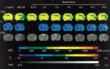
          Restoration of blood flow following ischemic stroke can be achieved by means of thrombolysis or mechanical recanalization. However, for some patients, reperfusion may exacerbate the injury initially caused by ischemia, producing a so-called “cerebral reperfusion injury”. Multiple pathological processes are involved in this injury, including leukocyte infiltration, platelet and complement activation, postischemic hyperperfusion, and breakdown of the blood–brain barrier.

          Methods/results and conclusions

          Magnetic resonance imaging (MRI) can provide extensive information on this process of injury, and may have a role in the future in stratifying patients’ risk for reperfusion injury following recanalization. Moreover, different MRI modalities can be used to investigate the various mechanisms of reperfusion injury. Antileukocyte antibodies, brain cooling and conditioned blood reperfusion are potential therapeutic strategies for lessening or eliminating reperfusion injury, and interventionalists may play a role in the future in using some of these therapies in combination with thrombolysis or embolectomy. The present review summarizes the mechanisms of reperfusion injury and focuses on the way each of those mechanisms can be evaluated by different MRI modalities. The potential therapeutic strategies are also discussed.

          Related collections

          Most cited references88

          • Record: found
          • Abstract: found
          • Article: not found

          Pathophysiology of ischaemia-reperfusion injury.

          Reperfusion of ischaemic tissues is often associated with microvascular dysfunction that is manifested as impaired endothelium-dependent dilation in arterioles, enhanced fluid filtration and leukocyte plugging in capillaries, and the trafficking of leukocytes and plasma protein extravasation in postcapillary venules. Activated endothelial cells in all segments of the microcirculation produce more oxygen radicals, but less nitric oxide, in the initial period following reperfusion. The resulting imbalance between superoxide and nitric oxide in endothelial cells leads to the production and release of inflammatory mediators (e.g. platelet-activating factor, tumour necrosis factor) and enhances the biosynthesis of adhesion molecules that mediate leukocyte-endothelial cell adhesion. Some of the known risk factors for cardiovascular disease (hypercholesterolaemia, hypertension, and diabetes) appear to exaggerate many of the microvascular alterations elicited by ischaemia and reperfusion (I/R). The inflammatory mediators released as a consequence of reperfusion also appear to activate endothelial cells in remote organs that are not exposed to the initial ischaemic insult. This distant response to I/R can result in leukocyte-dependent microvascular injury that is characteristic of the multiple organ dysfunction syndrome. Adaptational responses to I/R injury have been demonstrated that allow for protection of briefly ischaemic tissues against the harmful effects of subsequent, prolonged ischaemia, a phenomenon called ischaemic preconditioning. There are two temporally and mechanistically distinct types of protection afforded by this adaptational response, i.e. acute and delayed preconditioning. The factors (e.g. protein kinase C activation) that initiate the acute and delayed preconditioning responses appear to be similar; however the protective effects of acute preconditioning are protein synthesis-independent, while the effects of delayed preconditioning require protein synthesis. The published literature in this field of investigation suggests that there are several potential targets for therapeutic intervention against I/R-induced microvascular injury. Copyright 2000 John Wiley & Sons, Ltd.
            Bookmark
            • Record: found
            • Abstract: found
            • Article: not found

            Use of anti-ICAM-1 therapy in ischemic stroke: results of the Enlimomab Acute Stroke Trial.

            (2001)
            There has been recent interest in the possible role of reperfusion-induced inflammation with neuronal injury after stroke. Enlimomab, a murine intercellular adhesion molecule-1 (ICAM-1) antibody, reduces leukocyte adhesion and infarct size in experimental stroke studies. The purpose of the current clinical trial was to evaluate the use of enlimomab after ischemic stroke. A total of 625 patients with ischemic stroke were randomized to receive either enlimomab (n = 317) or placebo (n = 308) within 6 hours of stroke onset. Treatment was given over 5 days. Patients were evaluated at baseline and on days 5 and 90 after initiation of treatment; long-term assessments were carried out after 6 and 12 months. The primary efficacy endpoint was the response to therapy at 90 days on the Modified Rankin Scale; other endpoints included Barthel Index (BI) and NIH Stroke Scale and survival. At day 90, the Modified Rankin Scale score was worse in patients treated with enlimomab than with placebo (p = 0.004). Fewer patients had symptom-free recovery on enlimomab than placebo (p = 0.004), and more died (22.2 versus 16.2%). The negative effect of enlimomab was apparent on days 5, 30, and 90 of treatment (p = 0.005). There were significantly more adverse events with enlimomab treatment than placebo, primarily infections and fever. Patients experiencing fever were more likely to have a poor outcome or die. The authors conclude that anti-ICAM therapy with enlimomab is not an effective treatment for ischemic stroke in the model studied and, indeed, may significantly worsen stroke outcome.
              Bookmark
              • Record: found
              • Abstract: found
              • Article: not found

              Quantitative evaluation of blood-brain barrier permeability following middle cerebral artery occlusion in rats.

              A sensitive quantitative fluorescence method was used to explore the time course and regional pattern of blood-brain barrier (BBB) opening after transient middle cerebral artery occlusion (MCAo). Male Sprague-Dawley rats were anesthetized with halothane and subjected to 2 h of temporary MCAo by retrograde insertion of an intraluminal nylon suture, coated with poly-L-lysine, through the external carotid artery into the internal carotid artery and MCA. Damage to the BBB was judged by extravasation of Evans Blue (EB) dye, which was administered either 2, 3, 24 or 48 h after onset of MCAo. Fluorometric quantitation of EB was performed 1 or 2 h later in six brain regions. Cerebral infarction volumes were quantitated from histopathological material at 72 h. EB extravasation first became grossly visible in the ipsilateral caudoputamen and neocortex following 3 h of MCAo, was grossly unapparent at 24-26 h, and was maximal at 48-50 h. Fluorescence quantitation confirmed that BBB opening was absent at 2-3 h but present at all later times. In the hemisphere ipsilateral to MCAo, a 179% mean increase in extravasation of EB (compared to sham rats) was measured at 4 h, 407% at 5 h, 311% at 26 h and 264% at 50 h. (in each case, P < 0.05 vs. sham). The volume of infarcted tissue at 72 h in this model was 163.6 +/- 7.7 mm3. Our results indicate that an initial, acute disruption of the BBB occurs between 3 and 5 h following MCAo, and that a later, more widespread increase in regional BBB permeability is present at 48 h. Regional measurement of Evans Blue extravasation offers a precise means of quantitating BBB disruption in focal cerebral ischemia; this method will be of considerable utility in assessing the BBB-protective properties of pharmacological agents.
                Bookmark

                Author and article information

                Contributors
                ak2669@columbia.edu
                Journal
                Neuroradiology
                Neuroradiology
                Springer-Verlag (Berlin/Heidelberg )
                0028-3940
                1432-1920
                20 December 2006
                February 2007
                : 49
                : 2
                : 93-102
                Affiliations
                [1 ]Department of Radiology, Columbia University, 177 Fort Washington Ave, MHB 8SK, New York, NY 10032 USA
                [2 ]Pall Corporation, East Hills, NY USA
                Article
                183
                10.1007/s00234-006-0183-z
                1786189
                17177065
                4dc6b362-d503-4ff6-b013-ba966c091848
                © Springer-Verlag 2006
                History
                : 16 March 2006
                : 24 October 2006
                Categories
                Review
                Custom metadata
                © Springer-Verlag 2007

                Radiology & Imaging
                magnetic resonance imaging,hypothermia,conditioned blood reperfusion,cerebral ischemia,reperfusion injury

                Comments

                Comment on this article