87
views
0
recommends
+1 Recommend
0 collections
    0
    shares
      • Record: found
      • Abstract: found
      • Article: found
      Is Open Access

      Neutrophils with myeloid derived suppressor function deplete arginine and constrain T cell function in septic shock patients

      research-article

      Read this article at

      Bookmark
          There is no author summary for this article yet. Authors can add summaries to their articles on ScienceOpen to make them more accessible to a non-specialist audience.

          Abstract

          Introduction

          Impaired T cell function in sepsis is associated with poor outcome, but the mechanisms are unclear. In cancer, arginase-expressing myeloid derived suppressor cells (MDSCs) deplete arginine, impair T cell receptor CD3 zeta-chain expression and T cell function and are linked to poor clinical outcome, but their role during acute human infectious disease and in particular sepsis remains unknown. Hypoarginemia is prevalent in sepsis. This study aimed to determine whether neutrophils that co-purify with PBMC express arginase, and if arginine depletion constrains T cell CD3 zeta-chain expression and function in human sepsis.

          Methods

          Using flow cytometry, cell culture, HPLC, arginase activity and mRNA detection, our study examined whether neutrophils, with reduced buoyant density isolated in the Ficoll interface, metabolise L-arginine and suppress T cell proliferation in sepsis. A total of 35 sepsis patients (23 with septic shock) and 12 hospital controls in a tertiary referral hospital in tropical Australia were evaluated.

          Results

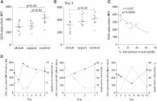
          Only sepsis patients had interphase neutrophils, neutrophils co-purifying with mononuclear cells (≤1.077 specific gravity). The percentage of interphase neutrophils in sepsis was proportional to sepsis severity and correlated with plasma IL-6 concentrations. Ex vivo, sepsis-derived interphase neutrophils expressed arginase, metabolised culture L-arginine and suppressed T cell proliferation and CD3 zeta-chain expression. In vivo, in septic shock there was a longitudinal inverse association between interphase neutrophil number and CD3 zeta-chain expression. Depletion or inhibition of interphase neutrophils in vitro restored zeta-chain expression and T cell function.

          Conclusions

          For the first time during an acute human infection, interphase neutrophils that express arginase were found to circulate in sepsis, in proportion to disease severity. These neutrophil-MDSCs impair T cell CD3 zeta-chain expression and T cell function via L-arginine metabolism, and likely contribute to the T cell dysfunction seen in sepsis. Modulation of neutrophil-MDSC or their downstream effects warrant consideration as targets for novel adjunctive therapies in sepsis.

          Electronic supplementary material

          The online version of this article (doi:10.1186/cc14003) contains supplementary material, which is available to authorized users.

          Related collections

          Most cited references42

          • Record: found
          • Abstract: found
          • Article: not found

          Activated granulocytes and granulocyte-derived hydrogen peroxide are the underlying mechanism of suppression of t-cell function in advanced cancer patients.

          Impaired T-cell function in patients with advanced cancer has been a widely acknowledged finding, but mechanisms reported thus far are those primarily operating in the tumor microenvironment. Very few mechanisms have been put forth to explain several well-described defects in peripheral blood T cells, such as reduction in expression of signaling molecules, decreased production of cytokines, or increased apoptosis. We have closely examined the peripheral blood mononuclear cell (PBMC) samples derived from patients and healthy individuals, and we have observed an important difference that may underlie the majority of reported defects. We observed that in samples from patients only, an unusually large number of granulocytes copurify with low density PBMCs on a density gradient rather than sediment, as expected, to the bottom of the gradient. We also show that activating granulocytes from a healthy donor with N-formyl-L-methionyl-L-leucyl-L-phenylalanine could also cause them to sediment aberrantly and copurify with PBMCs, suggesting that density change is a marker of their activation. To confirm this, we looked for other evidence of in vivo granulocyte activation and found it in drastically elevated plasma levels of 8-isoprostane, a product of lipid peroxidation and a marker of oxidative stress. Reduced T-cell receptor zeta chain expression and decreased cytokine production by patients' T cells correlated with the presence of activated granulocytes in their PBMCs. We showed that freshly obtained granulocytes from healthy donors, if activated, can also inhibit cytokine production by T cells. This action is abrogated by the addition of the hydrogen peroxide (H(2)O(2)) scavenger, catalase, implicating H(2)O(2) as the effector molecule. Indeed, when added alone, H(2)O(2) could suppress cytokine production of normal T cells. These findings indicate that granulocytes are activated in advanced cancer patients and that granulocyte-derived H(2)O(2) is the major cause of severe systemic T-cell suppression.
            Bookmark
            • Record: found
            • Abstract: found
            • Article: not found

            Characterization of cytokine-induced myeloid-derived suppressor cells from normal human peripheral blood mononuclear cells.

            Tumor immune tolerance can derive from the recruitment of suppressor cell populations, including myeloid-derived suppressor cells (MDSCs). In cancer patients, increased MDSCs correlate with more aggressive disease and a poor prognosis. Expression of 15 immune factors (TGFbeta, IL-1beta, IL-4, IL-6, IL-10, GM-CSF, M-CSF, IDO, fms-related tyrosine kinase 3 ligand, c-kit ligand, inducible NO synthase, arginase-1, TNF-alpha, cyclo-oxygenase 2, vascular endothelial growth factor [VEGF]) by MDSC-inducing human solid tumor cell lines was evaluated by RT-PCR. Based upon these data, cytokine mixtures were then tested for their ability to generate suppressive CD33(+) cells from healthy donor PBMCs in vitro by measuring their ability to inhibit the proliferation of, and IFN-gamma production by, fresh autologous human T cells after CD3/CD28 stimulation. Induced MDSCs were characterized with respect to their morphology, surface phenotype, and gene expression profile. MDSC-inducing cancer cell lines demonstrated multiple pathways for MDSC generation, including overexpression of IL-6, IL-1beta, cyclo-oxygenase 2, M-CSF, and IDO. CD33(+) cells with potent suppressive capacity were best generated in vitro by GM-CSF and IL-6, and secondarily by GM-CSF + IL-1beta, PGE(2), TNF-alpha, or VEGF. Characterization studies of cytokine-induced suppressive cells revealed CD33(+)CD11b(+)CD66b(+)HLA-DR(low)IL-13R alpha2(int) large mononuclear cells with abundant basophilic cytoplasm. Expression of inducible NO synthase, TGFbeta, NADPH oxidase, VEGF, and/or arginase-1 was also upregulated, and Transwell studies showed suppression of autologous T cells to be contact dependent. Suppressive CD33(+) cells generated from PBMCs by GM-CSF and IL-6 were consistent with human MDSCs. This study suggests that these cytokines are potential therapeutic targets for the inhibition of MDSC induction in cancer patients.
              Bookmark
              • Record: found
              • Abstract: found
              • Article: not found

              MyD88-dependent expansion of an immature GR-1+CD11b+ population induces T cell suppression and Th2 polarization in sepsis

              Polymicrobial sepsis alters the adaptive immune response and induces T cell suppression and Th2 immune polarization. We identify a GR-1+CD11b+ population whose numbers dramatically increase and remain elevated in the spleen, lymph nodes, and bone marrow during polymicrobial sepsis. Phenotypically, these cells are heterogeneous, immature, predominantly myeloid progenitors that express interleukin 10 and several other cytokines and chemokines. Splenic GR-1+ cells effectively suppress antigen-specific CD8+ T cell interferon (IFN) γ production but only modestly suppress antigen-specific and nonspecific CD4+ T cell proliferation. GR-1+ cell depletion in vivo prevents both the sepsis-induced augmentation of Th2 cell–dependent and depression of Th1 cell–dependent antibody production. Signaling through MyD88, but not Toll-like receptor 4, TIR domain–containing adaptor-inducing IFN-β, or the IFN-α/β receptor, is required for complete GR-1+CD11b+ expansion. GR-1+CD11b+ cells contribute to sepsis-induced T cell suppression and preferential Th2 polarization.
                Bookmark

                Author and article information

                Contributors
                christabelle.darcy@gmail.com
                gabriela.minigo@menzies.edu.au
                kim.piera@menzies.edu.au
                joshua.davis@menzies.edu.au
                yvette.mcneil@menzies.edu.au
                ywc@duke.edu
                avolk@duke.edu
                brice@duke.edu
                nicholas.anstey@menzies.edu.au
                Tonia.Woodberry@menzies.edu.au
                Journal
                Crit Care
                Critical Care
                BioMed Central (London )
                1364-8535
                1466-609X
                1 August 2014
                1 August 2014
                2014
                : 18
                : 4
                : R163
                Affiliations
                [ ]Global and Tropical Health Division, Menzies School of Health Research and Charles Darwin University, Casuarina NT 0811, P.O. Box 41096, Darwin, NT 0810 Australia
                [ ]Infectious Diseases Department, Royal Darwin Hospital, Darwin, NT 0810 Australia
                [ ]Division of Hematology-Oncology, Duke University and Veterans Affairs Medical Centers, Durham, NC 27705 USA
                Article
                2719
                10.1186/cc14003
                4261583
                25084831
                4ee4171d-1e5b-4ecb-b678-477e35ad3a53
                © Darcy et al.; licensee BioMed Central Ltd. 2014

                This article is published under license to BioMed Central Ltd. This is an Open Access article distributed under the terms of the Creative Commons Attribution License ( http://creativecommons.org/licenses/by/4.0), which permits unrestricted use, distribution, and reproduction in any medium, provided the original work is properly credited. The Creative Commons Public Domain Dedication waiver ( http://creativecommons.org/publicdomain/zero/1.0/) applies to the data made available in this article, unless otherwise stated.

                History
                : 24 February 2014
                : 18 June 2014
                Categories
                Research
                Custom metadata
                © The Author(s) 2014

                Emergency medicine & Trauma
                Emergency medicine & Trauma

                Comments

                Comment on this article