17
views
0
recommends
+1 Recommend
0 collections
    0
    shares
      • Record: found
      • Abstract: found
      • Article: found
      Is Open Access

      Oxidative Stress Induces Caveolin 1 Degradation and Impairs Caveolae Functions in Skeletal Muscle Cells

      research-article

      Read this article at

      Bookmark
          There is no author summary for this article yet. Authors can add summaries to their articles on ScienceOpen to make them more accessible to a non-specialist audience.

          Abstract

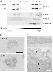
          Increased level of oxidative stress, a major actor of cellular aging, impairs the regenerative capacity of skeletal muscle and leads to the reduction in the number and size of muscle fibers causing sarcopenia. Caveolin 1 is the major component of caveolae, small membrane invaginations involved in signaling and endocytic trafficking. Their role has recently expanded to mechanosensing and to the regulation of oxidative stress-induced pathways. Here, we increased the amount of reactive oxidative species in myoblasts by addition of hydrogen peroxide (H 2O 2) at non-toxic concentrations. The expression level of caveolin 1 was significantly decreased as early as 10 min after 500 μM H 2O 2 treatment. This reduction was not observed in the presence of a proteasome inhibitor, suggesting that caveolin 1 was rapidly degraded by the proteasome. In spite of caveolin 1 decrease, caveolae were still able to assemble at the plasma membrane. Their functions however were significantly perturbed by oxidative stress. Endocytosis of a ceramide analog monitored by flow cytometry was significantly diminished after H 2O 2 treatment, indicating that oxidative stress impaired its selective internalization via caveolae. The contribution of caveolae to the plasma membrane reservoir has been monitored after osmotic cell swelling. H 2O 2 treatment increased membrane fragility revealing that treated cells were more sensitive to an acute mechanical stress. Altogether, our results indicate that H 2O 2 decreased caveolin 1 expression and impaired caveolae functions. These data give new insights on age-related deficiencies in skeletal muscle.

          Related collections

          Most cited references47

          • Record: found
          • Abstract: found
          • Article: not found

          Caveolae as plasma membrane sensors, protectors and organizers.

          Caveolae are submicroscopic, plasma membrane pits that are abundant in many mammalian cell types. The past few years have seen a quantum leap in our understanding of the formation, dynamics and functions of these enigmatic structures. Caveolae have now emerged as vital plasma membrane sensors that can respond to plasma membrane stresses and remodel the extracellular environment. Caveolae at the plasma membrane can be removed by endocytosis to regulate their surface density or can be disassembled and their structural components degraded. Coat proteins, called cavins, work together with caveolins to regulate the formation of caveolae but also have the potential to dynamically transmit signals that originate in caveolae to various cellular destinations. The importance of caveolae as protective elements in the plasma membrane, and as membrane organizers and sensors, is highlighted by links between caveolae dysfunction and human diseases, including muscular dystrophies and cancer.
            Bookmark
            • Record: found
            • Abstract: found
            • Article: not found

            Cells respond to mechanical stress by rapid disassembly of caveolae.

            The functions of caveolae, the characteristic plasma membrane invaginations, remain debated. Their abundance in cells experiencing mechanical stress led us to investigate their role in membrane-mediated mechanical response. Acute mechanical stress induced by osmotic swelling or by uniaxial stretching results in a rapid disappearance of caveolae, in a reduced caveolin/Cavin1 interaction, and in an increase of free caveolins at the plasma membrane. Tether-pulling force measurements in cells and in plasma membrane spheres demonstrate that caveola flattening and disassembly is the primary actin- and ATP-independent cell response that buffers membrane tension surges during mechanical stress. Conversely, stress release leads to complete caveola reassembly in an actin- and ATP-dependent process. The absence of a functional caveola reservoir in myotubes from muscular dystrophic patients enhanced membrane fragility under mechanical stress. Our findings support a new role for caveolae as a physiological membrane reservoir that quickly accommodates sudden and acute mechanical stresses. Copyright © 2011 Elsevier Inc. All rights reserved.
              Bookmark
              • Record: found
              • Abstract: found
              • Article: not found

              PTRF-Cavin, a conserved cytoplasmic protein required for caveola formation and function.

              Caveolae are abundant cell-surface organelles involved in lipid regulation and endocytosis. We used comparative proteomics to identify PTRF (also called Cav-p60, Cavin) as a putative caveolar coat protein. PTRF-Cavin selectively associates with mature caveolae at the plasma membrane but not Golgi-localized caveolin. In prostate cancer PC3 cells, and during development of zebrafish notochord, lack of PTRF-Cavin expression correlates with lack of caveolae, and caveolin resides on flat plasma membrane. Expression of PTRF-Cavin in PC3 cells is sufficient to cause formation of caveolae. Knockdown of PTRF-Cavin reduces caveolae density, both in mammalian cells and in the zebrafish. Caveolin remains on the plasma membrane in PTRF-Cavin knockdown cells but exhibits increased lateral mobility and accelerated lysosomal degradation. We conclude that PTRF-Cavin is required for caveola formation and sequestration of mobile caveolin into immobile caveolae.
                Bookmark

                Author and article information

                Contributors
                Role: Academic Editor
                Journal
                PLoS One
                PLoS ONE
                plos
                plosone
                PLoS ONE
                Public Library of Science (San Francisco, CA USA )
                1932-6203
                23 March 2015
                2015
                : 10
                : 3
                : e0122654
                Affiliations
                [1 ]Univ Bordeaux, Chimie et Biologie des Membranes et Nanoobjets, UMR 5248, F-33600 Pessac, France; CNRS, Chimie et Biologie des Membranes et Nanoobjets, UMR 5248, F-33600 Pessac, France; Bordeaux INP, Chimie et Biologie des Membranes et Nanoobjets, UMR 5248, F-33600 Pessac, France
                [2 ]Institut Curie—Centre de Recherche, Membrane Dynamics and Mechanics of Intracellular Signaling Team, INSERM U1143, CNRS UMR 3666, Paris, France
                Institut de Myologie, FRANCE
                Author notes

                Competing Interests: The authors have declared that no competing interests exist.

                Conceived and designed the experiments: AM SP CL OL ED MD. Performed the experiments: AM SP ED MD. Analyzed the data: AM SP ED OL. Contributed reagents/materials/analysis tools: AM SP MD ED. Wrote the paper: ED SP MD OL CL.

                Article
                PONE-D-14-33769
                10.1371/journal.pone.0122654
                4370508
                25799323
                4fac142d-d36e-408e-87d0-3bcd54cf8593
                Copyright @ 2015

                This is an open access article distributed under the terms of the Creative Commons Attribution License, which permits unrestricted use, distribution, and reproduction in any medium, provided the original author and source are credited

                History
                : 31 July 2014
                : 21 February 2015
                Page count
                Figures: 6, Tables: 3, Pages: 19
                Funding
                A PhD fellowship was awarded to Alexis Mougeolle by the region Aquitaine and the Institut Polytechnique Bordeaux (IPB) for three years (grant no. 20111301001). The Association Française contre les Myopathies (AFM) also supported this work during the last two years (grant no. 15485, name “CAVEOX”). C. Lamaze work on caveolae is supported by an AFM grant no. 15717 (“CAV-DYS-STRESS”). The funders had no role in study design, data collection and analysis, decision to publish, or preparation of the manuscript.
                Categories
                Research Article
                Custom metadata
                All relevant data are within the paper.

                Uncategorized
                Uncategorized

                Comments

                Comment on this article