55
views
0
recommends
+1 Recommend
0 collections
    0
    shares
      • Record: found
      • Abstract: found
      • Article: not found

      Katanin controls mitotic and meiotic spindle length

      research-article

      Read this article at

      ScienceOpenPublisherPMC
      Bookmark
          There is no author summary for this article yet. Authors can add summaries to their articles on ScienceOpen to make them more accessible to a non-specialist audience.

          Abstract

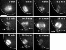
          Accurate control of spindle length is a conserved feature of eukaryotic cell division. Lengthening of mitotic spindles contributes to chromosome segregation and cytokinesis during mitosis in animals and fungi. In contrast, spindle shortening may contribute to conservation of egg cytoplasm during female meiosis. Katanin is a microtubule-severing enzyme that is concentrated at mitotic and meiotic spindle poles in animals. We show that inhibition of katanin slows the rate of spindle shortening in nocodazole-treated mammalian fibroblasts and in untreated Caenorhabditis elegans meiotic embryos. Wild-type C. elegans meiotic spindle shortening proceeds through an early katanin-independent phase marked by increasing microtubule density and a second, katanin-dependent phase that occurs after microtubule density stops increasing. In addition, double-mutant analysis indicated that γ-tubulin–dependent nucleation and microtubule severing may provide redundant mechanisms for increasing microtubule number during the early stages of meiotic spindle assembly.

          Related collections

          Most cited references41

          • Record: found
          • Abstract: found
          • Article: not found

          The bipolar mitotic kinesin Eg5 moves on both microtubules that it crosslinks.

          During cell division, mitotic spindles are assembled by microtubule-based motor proteins. The bipolar organization of spindles is essential for proper segregation of chromosomes, and requires plus-end-directed homotetrameric motor proteins of the widely conserved kinesin-5 (BimC) family. Hypotheses for bipolar spindle formation include the 'push-pull mitotic muscle' model, in which kinesin-5 and opposing motor proteins act between overlapping microtubules. However, the precise roles of kinesin-5 during this process are unknown. Here we show that the vertebrate kinesin-5 Eg5 drives the sliding of microtubules depending on their relative orientation. We found in controlled in vitro assays that Eg5 has the remarkable capability of simultaneously moving at approximately 20 nm s(-1) towards the plus-ends of each of the two microtubules it crosslinks. For anti-parallel microtubules, this results in relative sliding at approximately 40 nm s(-1), comparable to spindle pole separation rates in vivo. Furthermore, we found that Eg5 can tether microtubule plus-ends, suggesting an additional microtubule-binding mode for Eg5. Our results demonstrate how members of the kinesin-5 family are likely to function in mitosis, pushing apart interpolar microtubules as well as recruiting microtubules into bundles that are subsequently polarized by relative sliding.
            Bookmark
            • Record: found
            • Abstract: found
            • Article: not found

            On the control of oocyte meiotic maturation and ovulation in Caenorhabditis elegans.

            Prior to fertilization, oocytes undergo meiotic maturation (cell cycle progression) and ovulation (expulsion from the ovary). To begin the study of these processes in Caenorhabditis elegans, we have defined a time line of germline and somatic events by video microscopy. As the oocyte matures, its nuclear envelope breaks down and its cell cortex rearranges. Immediately thereafter, the oocyte is ovulated by increasing contraction of the myoepithelial gonadal sheath and relaxation of the distal spermatheca. By systematically altering the germ cell contents of the hermaphrodite using mutant strains, we have uncovered evidence of four cell-cell interactions that regulate maturation and ovulation. (1) Both spermatids and spermatozoa induce oocyte maturation. In animals with a feminized germline, maturation is inhibited and oocytes arrest in diakinesis. The introduction of sperm by mating restores maturation. (2) Sperm also directly promote sheath contraction. In animals with a feminized or tumorous germline, contractions are infrequent, whereas in animals with a masculinized germline or with sperm introduced by mating, contractions are frequent. (3 and 4) The maturing oocyte both induces spermathecal dilation and modulates sheath contractions at ovulation; dilation of the distal spermatheca and sharp increases in sheath contraction rates are only observed in the presence of a maturing oocyte. Copyright 1999 Academic Press.
              Bookmark
              • Record: found
              • Abstract: found
              • Article: not found

              Polarization of chemoattractant receptor signaling during neutrophil chemotaxis.

              Morphologic polarity is necessary for chemotaxis of mammalian cells. As a probe of intracellular signals responsible for this asymmetry, the pleckstrin homology domain of the AKT protein kinase (or protein kinase B), tagged with the green fluorescent protein (PHAKT-GFP), was expressed in neutrophils. Upon exposure of cells to chemoattractant, PHAKT-GFP is recruited selectively to membrane at the cell's leading edge, indicating an internal signaling gradient that is much steeper than that of the chemoattractant. Translocation of PHAKT-GFP is inhibited by toxin-B from Clostridium difficile, indicating that it requires activity of one or more Rho guanosine triphosphatases.
                Bookmark

                Author and article information

                Journal
                J Cell Biol
                jcb
                The Journal of Cell Biology
                The Rockefeller University Press
                0021-9525
                1540-8140
                18 December 2006
                : 175
                : 6
                : 881-891
                Affiliations
                [1 ]Section of Molecular and Cellular Biology, University of California, Davis, Davis, CA 95616
                [2 ]Department of Cellular and Molecular Medicine, Ludwig Institute for Cancer Research, University of California, San Diego, La Jolla, CA 92093
                Author notes

                Correspondence to Francis J. McNally: fjmcnally@ 123456ucdavis.edu

                Article
                200608117
                10.1083/jcb.200608117
                2064698
                17178907
                4fe145cf-833d-4c87-b2fa-334743eba661
                Copyright © 2006, The Rockefeller University Press
                History
                : 21 August 2006
                : 20 November 2006
                Categories
                Research Articles
                Article

                Cell biology
                Cell biology

                Comments

                Comment on this article