10
views
0
recommends
+1 Recommend
0 collections
    0
    shares
      • Record: found
      • Abstract: found
      • Article: found
      Is Open Access

      Bifid mandibular canal: Report of 2 cases and review of literature

      letter

      Read this article at

      Bookmark
          There is no author summary for this article yet. Authors can add summaries to their articles on ScienceOpen to make them more accessible to a non-specialist audience.

          Abstract

          Sir, The mandibular canal runs from the mandibular foramen to the mental foramen and contains the inferior alveolar artery, vein, and nerve. In medical imaging, its appearance has been described as “a radiolucent dark ribbon between two white lines.”[1] White and Pharoah defined it as “dark linear shadow with thin radiopaque superior and inferior borders cast by the lamella of bone that bounds the canal.”[2] Recognition of the mandibular canal variations is very important because of its clinical implications. 
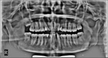
Here, we represent incidental findings of the same in our 2 cases [Figures 1 and 2]. Figure 1 Case 1 Figure 2 Case 2 The term “bifid” is derived from Latin, meaning a cleft into two parts or branches. Bifid mandibular canals originate at the mandibular foramen and might each contain a neurovascular bundle. The various types of bifid mandibular canals have been classified according to anatomical location and configuration. Smaller accessory canals might be seen in association with normal or bifid mandibular canals. Results of previous anatomical and radiological studies demonstrate significant variation in the course of the mandibular canal. According to Chávez-Lomeli et al., during embryologic development, the three inferior dental nerves innervating the three groups of mandibular teeth fuse together and form a single unified nerve in one canal. This theory would explain the existence of accessory canals resulting from lack of fusion of these canals.[3] In 1973, Kiersch and Jordan annotated that an osteocondensation image produced by the insertion of the mylohyoid muscle into the internal mandibular surface, with a distribution parallel to the dental canal, may mimic a bifid mandibular canal.[4] The imprint of the mylohyoid nerve on the internal mandibular surface, where it separates from the inferior alveolar nerve and travels to the floor of the mouth, may also be a cause for confusion.[5] A two-dimensional radiograph, such as a panoramic view, cannot completely rule out the possibility of a deep mylohyoid groove on the medial aspect of mandibular surface as the image on these two-dimensional representations can be confused with the second mandibular canal.[6] The incidence of bifid mandibular canal seems to be very low. Recently, there were two reports of bifid mandibular canals (6 cases in all) being diagnosed with the use of volumetric imaging (multislice helical computed tomography [CT] and cone-beam CT).[7] It seems that for accurate observation of the location and configuration of the mandibular canals, it is necessary to use cross-sectional images, taken perpendicular to the axis of the canals. However, CT scan, due to its high cost and radiation exposure, cannot be performed for all patients.[8] The clinical relevance of this issue is to remind clinicians of the variable anatomy of the mandibular canal. Inadequate anesthesia may be possible with any bifurcation type, but especially when there are two mandibular foramina. It may lead to complications while performing an inferior alveolar nerve block for obtaining mandibular anesthesia.[9] The location and configuration of mandibular canal variations have important implications in surgical procedures involving the mandible such as dental implant treatment, sagittal split ramus osteotomy, and orthognathic and reconstructive surgeries; displacement of the third molar into the nerve canal during surgery, bleeding, and traumatic neuroma are some of its other complications.[10] In patients wearing prostheses, this condition can cause pain and discomfort due to bone resorption. Using implants in these patients can also cause damage to the second canal. Therefore, it is of considerable interest for dentists to identify the presence of bifid canals on the panoramic radiographs to provide better patient care. Financial support and sponsorship Nil. Conflicts of interest There are no conflicts of interest.

          Related collections

          Most cited references10

          • Record: found
          • Abstract: found
          • Article: not found

          Observation of bifid mandibular canal using cone-beam computerized tomography.

          Some variations of the mandibular canal (so-called bifid mandibular canal) have been reported using various radiography techniques; however, the occurrences of bifid mandibular canal were less than 1% according to panoramic radiographic surveys. The purpose of the present investigation was to clarify the rate and type of bifid mandibular canal in the mandibular ramus region, as observed using cone-beam computerized tomography (CBCT) images. One hundred twenty-two patients who had undergone preoperative imaging of dental implant treatment using CBCT were enrolled in the investigation. Two-dimensional (2D) images of various planes in the mandibular ramus region were reconstructed on a computer using three-dimensional visualization and measurement software. The course of the mandibular canal was observed and the length of the bifid canal was measured. Bifid mandibular canal in the mandibular ramus region was observed in 65% of patients and 43% of sides. Bifid mandibular canal can be classified into four types: retromolar, dental, forward, and buccolingual canals. Bifid mandibular canal was observed at a high rate using CBCT.
            Bookmark
            • Record: found
            • Abstract: found
            • Article: not found

            The human mandibular canal arises from three separate canals innervating different tooth groups.

            The purpose of this study was to describe the prenatal formation of the human mandibular canal. Since bony canals develop in prenatal life around the nerve paths, it was assumed that the canal pattern could reflect the pattern of innervation of the dentition. Mapping of this early canal pattern does not appear to have been undertaken before. The material consisted of anthropological mandibles from the National Institute of Anthropology and History, Mexico City. A total of 302 human hemimandibles from the latter half of the prenatal period was investigated. The length, measured from the mental symphysis to the mandibular condyle, ranged from 28 to 60 mm. The dento-alveolar maturity was classified in two stages according to the appearance of alveolar sockets of deciduous and first permanent molars. The mandibles were radiographed with guttapercha points inserted into the canal openings (foramina) on the lingual surfaces of the mandibular rami. The study showed that the canal to the incisors appeared first, followed by the canal to the primary molars, and last by the one or more canals to the first permanent molars. In the most mature group, three different canals always occurred in each hemimandible. The canals were directed from the lingual surface of the mandibular ramus toward the different tooth groups. The inferior alveolar nerve presumably occurs in the mandible as three individual nerve paths originating at different stages of development. It is suggested that rapid prenatal growth and remodeling in the ramus region result in a gradual coalescence of the canal entrances that is obvious at birth. It is hypothesized that the pattern of tooth agenesis within the three groups of teeth is related to the three separate paths of innervation of the dentition.
              Bookmark
              • Record: found
              • Abstract: found
              • Article: not found

              Bifid mandibular canal in Japanese.

              The location and configuration of mandibular canal variations are important in surgical procedures involving the mandible, such as extraction of an impacted third molar, dental implant treatment, and sagittal split ramus osteotomy. We report 3 Japanese patients with bifid mandibular canals using panoramic radiograph and multi-slice helical computed tomography (CT) images. In 2 of the 5 sides, the bifid mandibular canal was suggested on panoramic radiograph. The bifid mandibular canal had a short and narrow upper canal toward the distal area of the second molar in 4 sides, and a short and narrow lower canal toward the distal area of second molar in 1 side, as revealed on reconstructed CT images. Since the location and configuration of mandibular canal variations are important in surgical procedures involving the mandible, they should be carefully observed using reconstructed CT images.
                Bookmark

                Author and article information

                Journal
                Saudi J Anaesth
                Saudi J Anaesth
                SJA
                Saudi Journal of Anaesthesia
                Medknow Publications & Media Pvt Ltd (India )
                1658-354X
                0975-3125
                Oct-Dec 2016
                : 10
                : 4
                : 488-489
                Affiliations
                [1]Department of Oral Medicine and Radiology, ACPM Dental College, Dhule, Maharashtra, India
                [1 ]Department of Oral Medicine and Radiology, M.G.V.'s K.B.H. Dental College and Hospital, Nashik, Maharashtra, India
                Author notes
                Address for correspondence: Dr. Ujwala A. Brahmankar, Department of Oral Medicine and Radiology, ACPM Dental College, Dhule - 424 003, Maharashtra, India. E-mail: drujwala.brahmankar@ 123456gmail.com
                Article
                SJA-10-488
                10.4103/1658-354X.179123
                5044749
                507baeb6-a055-4b22-8bb4-44139bce55b1
                Copyright: © Saudi Journal of Anaesthesia

                This is an open access article distributed under the terms of the Creative Commons Attribution-NonCommercial-ShareAlike 3.0 License, which allows others to remix, tweak, and build upon the work non-commercially, as long as the author is credited and the new creations are licensed under the identical terms.

                History
                Categories
                Letter to Editor

                Anesthesiology & Pain management
                Anesthesiology & Pain management

                Comments

                Comment on this article