54
views
0
recommends
+1 Recommend
0 collections
    0
    shares
      • Record: found
      • Abstract: found
      • Article: found
      Is Open Access

      Cystic Echinococcosis: Chronic, Complex, and Still Neglected

      research-article
      1 , 2 , 3 , * , 4 , on behalf of the members of the International CE Workshop in Lima, Peru, 2009
      PLoS Neglected Tropical Diseases
      Public Library of Science

      Read this article at

      Bookmark
          There is no author summary for this article yet. Authors can add summaries to their articles on ScienceOpen to make them more accessible to a non-specialist audience.

          Abstract

          The Overall Scene Cystic echinococcosis (CE), an infection with the larval form of the dog tapeworm Echinococcus granulosus, still causes serious lung and liver disease with a worldwide geographical distribution. This parasitic infection is preventable, eliminable, and treatable—in theory. The biological cycle can be attacked at various points: regular dog deworming, controlled sheep slaughtering, vaccination of the intermediate (sheep) animal host, and possibly in the future, vaccination of the definitive (dog) animal host (Figure 1). However, breaking the cycle in practice is difficult and requires long-lasting efforts. Control programs are expensive to set up and sustain. With the currently available options, a period of 20 years is needed to reach elimination, a goal that, unsurprisingly, has only been reached in rich countries [1]. 10.1371/journal.pntd.0001146.g001 Figure 1 Life cycle of Echinococcus granulosus in a community of the Middle Atlas region, Morocco. (We thank M. Kachani, College of Veterinary Medicine, Western University of Health Sciences, for the pictures.) At the current pace of control, patients suffering from CE will be seen for many decades to come. CE disease is chronic, complex, and neglected [2]–[4]. It is still poorly understood, and recommendations for diagnosis and treatment have not progressed beyond expert opinions and are not necessarily adopted by clinicians because of lack of grade I evidence. The critical issues are: 1. CE may develop silently over years and even decades until it surfaces with signs and symptoms or as a chance finding on an ultrasound (US) scan or chest X-rays requested for unrelated reasons. Clinical manifestations may mean that the cyst is already complicated, e.g., ruptured into the biliary or bronchial tree, secondarily infected with bacteria, or leaking and causing allergic reactions if not anaphylactic shock. 2. Screening large samples of populations to detect asymptomatic cases is expensive. As with all screening procedures, ethical issues arise: do all patients in whom cysts are found require treatment? Is the treatment which we then offer well established and safe? And is it available at all? Screening projects in endemic areas are often inadequately prepared, as the clinical management is not provided locally for those who are found positive. Problems start with the screening tool. With the exception of liver US, the available methods are far from satisfactory. In regards to serology, the sensitivity and specificity of several antigens have been well defined [5], [6], but available assays still lack standardization, sensitivity, and specificity [7]. Controversies on the usefulness for clinical diagnosis and screening remain unresolved [8]. Serodiagnostic performance depends on several factors, such as cyst location, cyst stage, and even cyst size, but these and other variables have not been thoroughly assessed to date. Ultrasound is an indispensable tool, but will likely miss very small cysts, and its efficacy is mostly restricted to intraabdominal organs. Additionally, some cyst stages may be difficult to distinguish from non-parasitic cysts, which are common. The problem continues when an echinococcal cyst has been diagnosed. In settings where health care facilities are several days of travel away from the rural areas where patients live and work, and as long as we have doubts on what the natural evolution of their cysts will be, clinical decision making is difficult. It has to be done in each case individually based on current standards, clinicians' experience, and local technical possibilities, supported by embarrassingly poor evidence. 3. Not all CE patients are similar, even at a population level. Broadly speaking, there are two defined groups of patients, each with a different set of problems: mainly asymptomatic patients (detected in screening programs or by chance), or clinically apparent cases (mostly patients with complicated cysts). (a) Patients with cysts detected during screening activities or as a chance finding. They mostly receive the treatment with which the attending clinician is familiar. This is not necessarily the best option relative to the cyst stage and clinical situation of the patient. Preliminary results from a survey on knowledge, attitudes, and practices regarding clinical management of CE in European, North African, and Middle Eastern countries yielded alarming results [9]. Patients may be put at risk of interventions that may be completely unnecessary. This certainly applies to a sizeable number of cysts that have become inactive and do not cause any symptoms or complications. A significant proportion of cysts stop growing and follow a path to spontaneous involution. Long-term follow-up suggests that these cysts and the patients harbouring them should be left alone. This is an appealing perspective for patients and health services, if evidence can be gathered in its support. CE4 and CE5 cysts appear to be very good candidates for this approach if they do not compromise any vital structures. It is, however, unclear if and under which circumstances this concept can be extended to other cyst types. (b) Patients developing complications. Successful management depends on equipment, skills, and quality of available health services. The most common complications are biliary obstruction with or without cholangitis, bronchial obstruction, bacterial infection of the cyst cavity with abscess formation, rupture with anaphylactic reactions that range from mild to lethal anaphylactic shock, secondary echinococcosis (growth of new cysts caused by seeding of protoscolices, generally in a cavity such as the peritoneal space) following spillage of fluid from a cyst that ruptured either spontaneously or because of a therapeutic maneuver, and impaired function of organs and blood vessels compressed by growing adjacent cysts (Figure 2). In most endemic countries, the required setup is only met in major cities a long way off from where patients experiencing complications live. 10.1371/journal.pntd.0001146.g002 Figure 2 Severe and life threatening complications of CE. (A) Biliary obstruction/obstructive cholangitis due to biliary fistulas. (B) Liver abscess formation due to secondary bacterial infection of cysts. (C) Cyst rupture (*) followed by anaphylaxis and secondary echinococcosis. (D) Cysts exerting pressure on vital neighbouring structures (e.g., liver veins resulting in Budd-Chiari Syndrome). (E) Embolism of the right pulmonary artery (arrow) caused by cardiac CE and vascular invasion. (F) CE infestation of the posterior wall of the left heart replacing the myocardial layer at the base of the heart. (We thank W. Hosch, Department of Radiology, and A. Stiehl, Department of Gastroenterology, University Hospital Heidelberg, for the images.). What Is Available Today to Diagnose and Treat CE Patients? Ultrasound is well established as a tool to diagnose, stage, and follow up CE cysts in the liver and other locations. Gharbi and colleagues developed the first widely adopted US classification in 1981 [10]. Other classifications were subsequently produced but were not as widely used. In 1994, the World Health Organization (WHO)-Informal Working Group started developing an international standardised US classification that could be universally applied to replace the plethora of classifications previously 
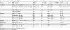
used (Figure 3) [11]. Even with all the obvious advantages of a standardised classification, some important issues still need to be resolved, one being the right sequence of cyst stages seen as the effect of natural or treatment-induced involution. A recent assessment of metabolic profiles of cyst stages with high-field proton magnetic resonance spectroscopy (1H MRS) has shown that the WHO IWGE classification of active, inactive, and transitional stages is perfectly in line with the metabolic activity profiles of the cysts, with the exception of CE3b, which appears vigorously active in 1H MRS, a finding that corresponds well with clinical experience [12]. US has been confirmed as an invaluable tool to assess cysts both with respect to viability and potential complications (Figure 2). 10.1371/journal.pntd.0001146.g003 Figure 3 Comparison of Gharbi's and WHO-IWGE ultrasound classifications of CE cysts. CL, as a potentially parasitic cyst, was not in Gharbi's classification and needs to be differentiated from non-parasitic cysts. This may also happen with CE1 cysts, when the double layer sign is not evident. Also, WHO CE3b had not been explicitly described by Gharbi but could likely be classified as Type III. There are basically four management options: surgery, percutaneous sterilization techniques, anti-parasitic treatment, and observation (“watch & wait”). Their individual roles were recently reviewed [2]–[4]. Each of the four strategies certainly has its place, but the specific places and boundaries are still not well defined. Surgery, the oldest form of treatment, keeps its place in most of the complicated forms of the disease. There is some competition between surgery and percutaneous approaches, in particular modified catheterization techniques, to be resolved, but this comparison requires carefully designed studies and cannot be decided on the basis of exclusively non-comparative small clinical studies, which are the only ones currently available. Proponents of classical PAIR (punction, aspiration, injection, reaspiration) [13] have lost a bit of their enthusiasm after realizing that some cyst stages, such as CE2 and CE3b, are quite tedious to needle with too many compartments to be individually approached. But most importantly, these stages tend to relapse after PAIR. It remains to be seen whether large modified catheterization techniques can substitute for PAIR in these stages. Over the past decade, several studies have been published suggesting that medical therapy (mebendazole, albendazole) could be an alternative to invasive treatment options in patients with uncomplicated cysts, broadening the indication for medical treatment over the years. The individual studies were all small and heterogeneity precluded appropriate meta-analysis. A recently published pooled analysis of individual patient data collected from six treatment centres suggests that the overall efficacy of benzimidazoles has been overrated [14]. Clinical trials stratified by cyst stage are needed to define the place of anti-parasitic treatment in the treatment of CE since it appears that it works better in some cyst stages (e.g., small CE1 cysts) than in others. The rate and nature of side effects of prolonged application of benzimidazole also deserves to be investigated more rigorously. Other anthelmintics, old and new (praziquantel, nitazoxanide), and combinations of anthelmintics (e.g., albendazole plus praziquantel) need to be properly investigated, too. Though so far not systematically studied, experience with leaving certain cysts completely alone and only following them up over years, points to a fourth managing option, watch & wait. Apart from being biologically plausible, long term follow-up of patients with CE4 and CE5 cysts in anatomically silent corners of the body looks good. This holds great promise for patients in whom cysts have reached this stage and needs to be urgently systematically studied. Reasons for Arrested Progress in CE Difficult, chronic diseases with a low case fatality rate clustering in poor rural areas are particularly “unattractive” to researchers and funders who depend on quick results to maintain the momentum of their activities. CE shares this fate with other communicable diseases, such as neurocysticercosis and Buruli disease. Health services also turn a blind eye on them since they plainly lack the means to manage patients with complex diseases such as CE appropriately. This is reflected in the low attention national and international institutions are paying to CE despite its substantial global burden, which is estimated at over 1 million DALYs per year [15], [16]. Additionally, due to its global distribution pattern, CE is not taking advantage of the attention that is being paid to “tropical” diseases. Interestingly, CE never made it to the list of the “TDR diseases” (from the WHO Special Programme for Research and Training in Tropical Diseases). The scarcity of resources and lack of momentum leads research to develop in niches with research communities too small to plan and conduct projects on a scale that allows conclusive answering of the relevant questions on efficacy, effectiveness, adverse reactions, and costs of a given treatment in comparison to other options. Currently available data arise from a multitude of small underpowered studies carried out over years, leading to contradicting results and recommendations, and, consequently, to controversies and difficulties (e.g., randomization) when planning appropriately designed clinical trials. What Do We Need to Improve CE Management in the Short Term? Here is a most clinically neglected parasitic disease that urgently needs attention. A valuable tool for diagnosing, staging, and following up patients, ultrasound, is readily available. Four management procedures, surgery, percutaneous sterilization techniques, anti-parasitic treatment, and watch & wait, have “evolved” over decades, and been recently summarized [4], but without adequate comparative evaluation of efficacy, effectiveness, rate of adverse events, relapse rates, and cost. Clinical decision making is on even shakier ground for extrahepatic and extrapulmonary locations, which are rarer (see [4] for a list of extrahepatic and extrapulmonary locations with related treatments), and numbers needed to build comparative trials hard to come by. There is an obligation to put at least what we have on an appropriate evidence base by conducting comparative clinical trials at the scale and quality that allow answering these important questions. As one of the expected results, clear criteria for the watch & wait option alone might already save a substantial proportion of patients from unnecessary interventions and save health services money. Difficult chronic diseases clustering in poor rural areas need intelligent, creative approaches, and this one urgently needs operational research incorporating the particularities of resource-poor settings into consideration.

          Related collections

          Most cited references15

          • Record: found
          • Abstract: found
          • Article: not found

          Ultrasound examination of the hydatic liver.

          Hydatic cysts of the liver were detected by ultrasound in 121 cases; all cases were confirmed surgically. Scans were classified based on sonographic analysis of the morphology and structure of the cyst. Five categories were found, which are thought to correspond to evolutionary stages of the hydatic cyst.
            Bookmark
            • Record: found
            • Abstract: found
            • Article: not found

            Clinical management of cystic echinococcosis: state of the art, problems, and perspectives.

            Clinical management of cystic echinococcosis (CE) has evolved over decades without adequate evaluation of important features such as efficacy, effectiveness, rate of adverse reactions, relapse rate, and cost. CE occurs in health care environments as different as Europe/North America and resource-poor countries of the South and the East. This creates setting-specific problems in the management of patients. Furthermore, studies carried out in either of the two fundamentally different environments lack external validity, i.e., results obtained in one setting may be different from those in the other and practices that can work in one may not be applicable to the other. In this paper, we review the current management procedures of CE with particular emphasis on the evidence base and setting-specific problems.
              Bookmark
              • Record: found
              • Abstract: found
              • Article: found
              Is Open Access

              Treatment Response of Cystic Echinococcosis to Benzimidazoles: A Systematic Review

              Introduction Cystic echinococcosis (CE, hydatid disease) is a parasitic disease of worldwide prevalence. Hydatid cysts occur mainly in the liver (70%) and the lung (20%). Clinical symptoms and signs depend on their localisation, size, and number. Currently four treatment modalities are in use: (1) surgery, (2) PAIR (puncture, aspiration, injection of protoscolicidal agent, reaspiration), (3) chemotherapy with albendazole (ABZ) or mebendazole (MBZ), and (4) watch and wait for inactive, clinically silent cysts. The evidence supporting any of the four treatment modalities, from carefully designed clinical studies, is insufficient, and choosing treatment options for patients remains controversial [1]. The use of benzimidazoles in CE treatment started in the 1970s with MBZ. In the early 1980s ABZ was introduced and since then has largely replaced mebendazole. The main advantages of ABZ are claimed to be a lower dosage and better intestinal absorption. In treatment centres MBZ and ABZ are given at the World Health Organisation (WHO) recommended dosages of (MBZ, 40–50 mg/kg/day; ABZ, 10–15 mg/kg/day) [2]. Variability exists in the duration of treatment, which remains undefined. Duration of treatment is determined according to the ultrasound-based treatment response, resulting in repetitive treatment, which is part of our analysis. Chemotherapy for the treatment of CE was initially recommended for inoperable patients and patients with multiple organ disease [2],[3]. Over the past decade several studies, mainly case series, have been published suggesting that chemotherapy could be an alternative to surgery in patients with uncomplicated cysts, leading to an increased use of chemotherapy over the years [4]. After more than 30 y of benzimidazole use, the following crucial question remains unanswered: what is the efficacy of benzimidazoles stratified by type and size of cysts, daily dose, and duration of treatment? This project started with a systematic review of the published literature on the efficacy of treating CE with benzimidazoles. We had to conclude, however, that we could not obtain a clear picture of the long-term outcome of benzimidazole treatment because inclusion criteria, treatment, outcome measures, and follow-up of published studies varied widely with substantial overlap of cohorts [1], thus precluding a meta-analysis of published results. We therefore initiated EchinoMEDREV, a collaborative effort of CE specialists, to collect individual patient data from patients treated with benzimidazoles. The main objectives of this collaborative study were to describe cyst outcome after initiation of benzimidazole treatment, with outcome defined by cyst stage determined by ultrasound following the WHO classification of 2001 [3], and to explore differences in outcome by cyst stage and size at initiation of treatment by using a common analytical strategy for all data across treatment centres. Methods Literature Search A systematic search of MEDLINE, EMBASE, CCTR, and SIGLE was carried out from their inception until week 4 of 2004. The search was performed by a research librarian using the following search terms: echinococcosis, albendazole, mebendazole, hydatid disease, cystic echinococcosis. We also searched reference lists and asked researchers in the field for additional studies. No language restriction was used. Abstracts were screened for suitability by MS. The eligibility of studies was assessed independently by two investigators (TJ and MS). We included all types of study design with a minimum of 30 patients treated either with ABZ or MBZ. Studies in which drug treatment was an adjunct to surgery, PAIR, or a second drug were excluded. Individual Patient and Cyst Data Collection The studies identified in the literature search revealed that there were large differences in baseline assessment of cyst stages, interventions (dose and duration of chemotherapy), length of follow-up, and outcome measures between published trials. These differences precluded the possibility to perform a meta-analysis of published results. Therefore we decided to collect individual patient data from the identified centres and initiated the EchinoMEDREV project. Study centres that had conducted clinical studies on benzimidazole treatment of CE were contacted and asked to contribute published and unpublished individual patient data of benzimidazole-treated CE patients. Data extraction forms were developed, piloted, and revised. Data collection started in June 2005 and ended in December 2007. Data were extracted from patient charts at the individual treatment centres. Data collected were: demographic data (age, sex); treatment data (MBZ, ABZ, dosage, and duration of treatment, side effects, previous treatments); imaging data (initial ultrasound staging and staging at follow-up visits). The forms were sent to the coordinating centre at the University Hospital in Heidelberg where data were electronically entered into a database with EpiData, using data entry checks. Accuracy in data entry was checked by double entry verification. A final dataset was created after correcting detected data entry errors and exported to Stata for statistical analysis. Patients with single or multiple hydatid cysts were eligible. Cyst stage had to be recorded at the beginning and at least once after completion of the initial treatment episode. The minimum follow-up period was 1 y after completion of initial treatment. Cyst activity had to be assessed by ultrasonography and classified according to WHO (CL–CE5 or active [A]/transitional [T]/inactive [I]), Gharbi, Perdomo, or Caremani (Table 1). 10.1371/journal.pntd.0000524.t001 Table 1 Ultrasound classification systems of CE cysts. Gharbi et al. 1981 [32] Perdomo et al. 1995 [33] Caremani et al. 1997 [34] WHO 2001 G I, G III P 1P 1a, 1b, 1cP 2 C IaC IbC IIa, IIb CLCE1CE2 A, active: unilocular simple cyst with uniform anechoic content with or without visible cyst wall or multivesicular, multiseptated cyst, presence of daughter cysts, round or oval G II, G IV P 3 C IIIa, IIIb; C IV CE3 T, transitional: unilocular cyst may contain daughter cysts, anechoic content detachment of membrane, form may be less rounded G V P 4, 4aP 5, 6 C Va, VbC VI, VIIa,VII b CE4CE5 I, inactive: heterogenous, hypoechoic, or hyperechoic degenerative content, no daughter cysts, calcification of cyst Data Analysis The analysis presented here includes only liver and peritoneal cysts (70%–75% of all CE cysts in humans), which were assessed by ultrasonography, and excluded lung cysts as they are not usually assessed by ultrasonography. The cyst was used as the unit of analysis for a description of achieved outcomes, and the presence of multiple cysts was not taken into account. Data were analysed by intention-to-continue-treatment, ignoring treatment changes (MBZ/ABZ), interruptions, and subsequent treatment episodes. We analysed several endpoints. First, initial treatment success for a cyst was defined as transformation from an initially active or transitional stage to an inactive stage or disappearance of the cyst (see Table 1 for classification based on ultrasonography). For this analysis time was measured from the start of first documented treatment to the date the stage was assessed as inactive or as disappeared or to the last documented assessment. Second, an analysis was made of the time for a cyst to become active again after the cysts had been staged as inactive; a necessary step, as some cysts that had reached an inactive stage had subsequently been staged as active again upon ultrasonography. For this analysis time was measured from the first (or second, or third) date at which a cyst was staged as inactive until the cyst was staged again as active. For these separate endpoints we performed time-to-event analyses using the Kaplan-Meier method and calculated the cumulative incidence of the events by subtracting the Kaplan-Meier survival estimate from one. Descriptive figures are presented stratified by centre where appropriate. Despite the fact that all previous studies on CE cyst development had treated cysts as an independent unit of analysis even if multiple cysts were present in the same patient, we addressed clustering of cysts within patients. The question of heterogeneity by centre was also examined with data from several treatment centres. When addressing clearly specified hypotheses—such as the association of cyst size and time to inactivity or cyst disappearance—Cox proportional hazards models were fitted and a robust variance estimator was used [5] to account for the clustering of cysts within patients. In addition indicator variables were included for each centre. Two questions were investigated using robust Cox proportional hazards model:(1) the association of cyst CE stage at baseline with time to first inactivity or disappearance, and (2) the association of cyst size ( 6 cm) with time to inactivity or cyst disappearance. For the second question the first year (day 0 to day 365) and the follow-up time after year one (day 366 onwards) were analysed separately, because descriptive cumulative incidence plots hinted at the possibility that the cyst size mattered only after year one. Wald test-based p-values were calculated to obtain a hypothesis test for a whole group of indicator variables to be included in the robust Cox proportional hazards model. p 6 cm. In the robust Cox proportional hazards model cysts <6 cm responded better than larger cysts (p = 0.006) and a strong centre effect was noted (p<0.0001). 10.1371/journal.pntd.0000524.g004 Figure 4 First time a cyst was staged as inactive or disappeared by size of cyst. Figure 5 shows the cumulative incidence of reaching an inactive stage or a disappearance of cysts for the first time by centres. Data from Greece and Bulgaria show inactivation/disappearance rates of cysts of 75%, increasing to around 90% within 2 y in Bulgaria. In contrast data from Palermo show inactivation or disappearance of cysts in approximately 20% of cases after 2 y. Data from Rome, Romania, and Turkey are between Greece and Palermo. 10.1371/journal.pntd.0000524.g005 Figure 5 First time a cyst was staged as inactive or disappeared by centre. Overall, cysts that reached an inactive stage for the first time relapsed (returned into an A or T stage) in around 25% of cases 2 y after inactivation (Figure 6). Cysts that reached an inactive stage for a second or third time showed relapse at a higher proportion and at an earlier stage: 60% of cysts relapsed within 2 y after the second or third inactivation. 10.1371/journal.pntd.0000524.g006 Figure 6 Time an active or transitional stage was reached stratified by the number of times an inactive stage had been reached previously (only centres that recorded recurrences are included). Figure 7 shows the proportion of inactive/disappeared cysts over time stratified by the first, second, and third time the cysts started from A/T stages. The cumulative incidence curve after first A/I reflects what has been observed after treatment initiation. Cysts that were staged A/T for the second and third time were staged as inactive or as disappeared in about 75% and 85% 1 y later. These results were almost exclusively from the Rome centre. 10.1371/journal.pntd.0000524.g007 Figure 7 Time an inactive or disappeared stage was reached for the next time, stratified by the number of times an active or transitional stage had been reached previously. In the simulation of hypothetical cysts, we estimated that 1 and 2 y after treatment initiation, 60% and 40% of cysts are still active or become active again. Discussion In a collaborative effort, individual data from patients with CE were pooled from six centres in five countries and outcomes of liver and peritoneal cysts treated with benzimidazoles were described. We found a strong association between cyst activity and response to treatment, with a better response in highly active CE1 cysts, and an association in treatment response depending on the size of the cyst at the beginning of treatment, with cysts <6 cm in diameter responding better. Thus, our data suggest that small highly active cysts show the best initial treatment response. Overall 25% of cysts reverted to active status within 1.5 to 2 y after having initially responded, and multiple relapses were observed. We estimated that 2 y after treatment initiation 40% of cysts are still active or become active again. Our results are biologically plausible because early in the disease host response resulting in an increasing thickness of the pericyst and consolidation of cyst content has not yet reached a degree that prevents the drug to reach its target [29]. Additionally, it is important to note that natural decay is a component of the observed rate of inactivation. Available data suggest that this decay may be as high as 13.6% within 18 mo [29], and up to 20.6% within 44 mo [30]. This finding clearly leads to an overestimation of response to benzimidazole treatment as calculated from longitudinal data, which increase with the length of observation. There are several limitations to this study. The published data collected from participating specialist centres are from case series. Results from case series are considered low level evidence in determining the efficacy of treatment options. In several analyses we found heterogeneity by centre. For example, the cumulative incidence curves for reaching an inactive cyst stage for the first time or the disappearance of a cyst after initial treatment showed large intercentre variability. Greek and Bulgarian data show a very rapid response, whereas data from Palermo show a very sluggish response to treatment. Time to first inactivation of cysts in the other centres looks quite similar. Rapid response to treatment in Greece and Bulgaria, however, remains unexplained. The particularly slow response to treatment shown in the dataset from Palermo could be due to the larger mean size of cysts at presentation, the difficulty of translating the Caremani classification into the WHO ultrasound classification (Table 1), and the well-known fact that inter-rater agreement between experts on classification of certain cyst stages is low, in particular for cysts containing daughter cysts—stage CE2 and stage CE3b according to WHO. Depending on the amount of consolidated matrix, cysts are either classified CE2 (daughter cysts with no matrix) or CE3b (daughter cysts with matrix).This distinction is important since the former is regarded active, the latter transitional [31]. Interobserver discrepancies occurred in the description of transitions from the inactive stages CE4 and CE5 to the active stages CE1 or CE2; some observers described these transitions and regard them as possible, whereas others do not. However, the number of misclassified cysts is not quantifiable in a retrospective dataset. It is very difficult, if not impossible, to consider all types and directions of biases when attempting to estimate the response of CE cysts to benzimidazoles from the available data. A very strong bias is certainly introduced by differing observation times with considerable impact on inactivation due to spontaneous involution. A final problem concerns data that we were unable to obtain (Table 2). Two centres that initially offered to deliver large datasets were eventually unable to do so. Two-thirds of our data have been provided by Rome, consequently the results are predominantly from one centre. Despite these limitations, to our knowledge, this study represents the largest CE dataset ever collected and analyzed in a uniform approach; further it is likely the only dataset obtained from the main international specialist groups. The recommendations on benzimidazole treatment of CE are currently based on the published results from these centres. Through the collection of individual patient data and the pooled analysis of these data we have managed to overcome some of the existing limitations present in the published literature. Does our study provide sufficient evidence to influence decisions for the treatment of CE? We think that our results are strong enough to cast doubts on overoptimistic expectations of the overall efficacy of benzimidazoles. When looking into substrata of the cyst population small CE1 cysts (diameter <6 cm) are a promising target for benzimidazole therapy, whereas stage CE2 and CE3 cysts respond poorly. The available evidence from this and other studies does not yet allow us, however, to formulate solid evidence-based drug treatment recommendations across all cyst stages, sizes, and locations. Our results highlight the urgent need to compare in a pragmatic randomised controlled trial the effect of standardized benzimidazole dose regimens on the individual active cyst stages (CE1, CE2, CE3a, and CE3b) substratified by cyst size. Such a trial would investigate as a primary outcome the proportion of cysts that become inactive (cyst stages CE4 and CE5) after treatment, and as a secondary outcome the yearly relapse rates up to 5 y after completion of treatment. The clarification of the efficacy of benzimidazoles in CE treatment is of paramount importance since benzimidazoles are the only drugs currently available to treat this neglected disease. Surgery as an alternative to benzimidazoles carries a significantly higher risk of adverse events, in particular intra- and postoperative morbidity and mortality and disseminated disease due to intraoperative spillage of viable hydatid material. Percutaneous fine needle techniques such as PAIR are only applicable to cyst stages CE1 and possibly CE3a, but not to CE2 and CE3b, which makes it necessary to explore large bore catheter techniques if albendazole turns out to be less effective in these cyst stages as suggested by our analysis. Supporting Information Checklist S1 QUOROM checklist. (0.17 MB PDF) Click here for additional data file.
                Bookmark

                Author and article information

                Contributors
                Role: Editor
                Journal
                PLoS Negl Trop Dis
                plos
                plosntds
                PLoS Neglected Tropical Diseases
                Public Library of Science (San Francisco, USA )
                1935-2727
                1935-2735
                July 2011
                26 July 2011
                : 5
                : 7
                : e1146
                Affiliations
                [1 ]Division of Infectious and Tropical Diseases, University of Pavia, IRCCS S.Matteo Hospital Foundation, WHO Collaborating Centre on Clinical Management of Cystic Echinococcosis, Pavia, Italy
                [2 ]Cysticercosis Unit, Instituto Nacional de Ciencias Neurológicas, Lima, Peru
                [3 ]Department of Microbiology, School of Sciences, and Center for Global Health, Universidad Peruana Cayetano Heredia, Lima, Peru
                [4 ]Section Clinical Tropical Medicine, Department of Infectious Diseases, University Hospital, Heidelberg, Germany
                New York Blood Center, United States of America
                Author notes

                ¶ Members of the International CE Workshop, listed in the Acknowledgments, actively participated in the development of the manuscript ideas and text.

                Article
                PNTD-D-10-00251
                10.1371/journal.pntd.0001146
                3144181
                21814584
                50ccfe1f-847e-48b2-b33e-880d297ab4b3
                Brunetti et al. This is an open-access article distributed under the terms of the Creative Commons Attribution License, which permits unrestricted use, distribution, and reproduction in any medium, provided the original author and source are credited.
                History
                Page count
                Pages: 5
                Categories
                Viewpoints
                Medicine
                Global Health
                Infectious Diseases
                Public Health
                Veterinary Science

                Infectious disease & Microbiology
                Infectious disease & Microbiology

                Comments

                Comment on this article