28
views
0
recommends
+1 Recommend
1 collections
    0
    shares

      Drug Design, Development and Therapy (submit here)

      This international, peer-reviewed Open Access journal by Dove Medical Press focuses on the design and development of drugs, as well as the clinical outcomes, patient safety, and programs targeted at the effective and safe use of medicines. Sign up for email alerts here.

      88,007 Monthly downloads/views I 4.319 Impact Factor I 6.6 CiteScore I 1.12 Source Normalized Impact per Paper (SNIP) I 0.784 Scimago Journal & Country Rank (SJR)


      • Record: found
      • Abstract: found
      • Article: found
      Is Open Access

      Cisplatin induces protective autophagy through activation of BECN1 in human bladder cancer cells

      research-article

      Read this article at

      ScienceOpenPublisherPMC
      Bookmark
          There is no author summary for this article yet. Authors can add summaries to their articles on ScienceOpen to make them more accessible to a non-specialist audience.

          Abstract

          Purpose

          Cisplatin-based chemotherapy is the first line treatment for several cancers including bladder cancer (BC). Autophagy induction has been implied to contribute to cisplatin resistance in ovarian cancer; and a high basal level of autophagy has been demonstrated in human bladder tumors. Therefore, it is reasonable to speculate that autophagy may account for the failure of cisplatin single treatment in BC. This study investigated whether cisplatin induces autophagy and the mechanism involved using human BC cell lines.

          Materials and methods

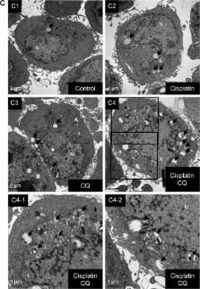
          Human BC cells (5637 and T24) were used in this study. Cell viability was detected using water soluble tetrazolium-8 reagents. Autophagy induction was detected by monitoring the levels of light chain 3 (LC3)-II and p62 by Western blot, LC3-positive puncta formation by immunofluorescence, and direct observation of the autophagolysosome (AL) formation by transmission electron microscopy. Inhibitors including bafilomycin A1 (Baf A1), chloroquine (CQ), and shRNA-based lentivirus against autophagy-related genes (ATG7 and ATG12) were utilized. Apoptosis level was detected by caspase 3/7 activity and DNA fragmentation.

          Results

          Cisplatin decreased cell viability and induced apoptosis of 5637 and T24 cells in a dose-and time-dependent manner. The increased LC3-II accumulation, p62 clearance, the number of LC3-positive puncta, and ALs in cisplatin-treated cells suggested that cisplatin indeed induces autophagy. Inhibition of cisplatin-induced autophagy using Baf A1, CQ, or ATG7/ATG12 shRNAs significantly enhanced cytotoxicity of cisplatin toward BC cells. These results indicated that cisplatin induced protective autophagy which may contribute to the development of cisplatin resistance and resulted in treatment failure. Mechanistically, upregulation of beclin-1 (BECN1) was detected in cisplatin-treated cells, and knockdown of BECN1 using shRNA attenuated cisplatin-induced autophagy and subsequently enhanced cisplatin-induced apoptosis.

          Conclusion

          Collectively, the study results indicated that cisplatin-induced autophagy is mediated by BECN1 in BC cells. Therefore, combinative treatment using cisplatin and autophagy inhibitors could potentially overcome cisplatin resistance related to autophagy induction.

          Most cited references29

          • Record: found
          • Abstract: found
          • Article: not found

          The role of autophagy in cancer development and response to therapy.

          Autophagy is a process in which subcellular membranes undergo dynamic morphological changes that lead to the degradation of cellular proteins and cytoplasmic organelles. This process is an important cellular response to stress or starvation. Many studies have shed light on the importance of autophagy in cancer, but it is still unclear whether autophagy suppresses tumorigenesis or provides cancer cells with a rescue mechanism under unfavourable conditions. What is the present state of our knowledge about the role of autophagy in cancer development, and in response to therapy? And how can the autophagic process be manipulated to improve anticancer therapeutics?
            Bookmark
            • Record: found
            • Abstract: found
            • Article: not found

            Dual role of 3-methyladenine in modulation of autophagy via different temporal patterns of inhibition on class I and III phosphoinositide 3-kinase.

            A group of phosphoinositide 3-kinase (PI3K) inhibitors, such as 3-methyladenine (3-MA) and wortmannin, have been widely used as autophagy inhibitors based on their inhibitory effect on class III PI3K activity, which is known to be essential for induction of autophagy. In this study, we systematically examined and compared the effects of these two inhibitors on autophagy under both nutrient-rich and deprivation conditions. To our surprise, 3-MA is found to promote autophagy flux when treated under nutrient-rich conditions with a prolonged period of treatment, whereas it is still capable of suppressing starvation-induced autophagy. We first observed that there are marked increases of the autophagic markers in cells treated with 3-MA in full medium for a prolonged period of time (up to 9 h). Second, we provide convincing evidence that the increase of autophagic markers is the result of enhanced autophagic flux, not due to suppression of maturation of autophagosomes or lysosomal function. More importantly, we found that the autophagy promotion activity of 3-MA is due to its differential temporal effects on class I and class III PI3K; 3-MA blocks class I PI3K persistently, whereas its suppressive effect on class III PI3K is transient. Because 3-MA has been widely used as an autophagy inhibitor in the literature, understanding the dual role of 3-MA in autophagy thus suggests that caution should be exercised in the application of 3-MA in autophagy study.
              Bookmark
              • Record: found
              • Abstract: found
              • Article: not found

              Application and interpretation of current autophagy inhibitors and activators

              Autophagy is the major intracellular degradation system, by which cytoplasmic materials are delivered to and degraded in the lysosome. As a quality control mechanism for cytoplasmic proteins and organelles, autophagy plays important roles in a variety of human diseases, including neurodegenerative diseases, cancer, cardiovascular disease, diabetes and infectious and inflammatory diseases. The discovery of ATG genes and the dissection of the signaling pathways involved in regulating autophagy have greatly enriched our knowledge on the occurrence and development of this lysosomal degradation pathway. In addition to its role in degradation, autophagy may also promote a type of programmed cell death that is different from apoptosis, termed type II programmed cell death. Owing to the dual roles of autophagy in cell death and the specificity of diseases, the exact mechanisms of autophagy in various diseases require more investigation. The application of autophagy inhibitors and activators will help us understand the regulation of autophagy in human diseases, and provide insight into the use of autophagy-targeted drugs. In this review, we summarize the latest research on autophagy inhibitors and activators and discuss the possibility of their application in human disease therapy.
                Bookmark

                Author and article information

                Journal
                Drug Des Devel Ther
                Drug Des Devel Ther
                Drug Design, Development and Therapy
                Dove Medical Press
                1177-8881
                2017
                16 May 2017
                : 11
                : 1517-1533
                Affiliations
                [1 ]Central Laboratory, Shin Kong Wu Ho-Su Memorial Hospital, Taipei
                [2 ]Division of Urology, School of Medicine, Fu-Jen Catholic University, New Taipei
                [3 ]Division of Urology, Department of Surgery, Shin Kong Wu Ho-Su Memorial Hospital
                [4 ]Department of Urology, Taipei Medical University, Taipei, Taiwan
                Author notes
                Correspondence: Thomas I-Sheng Hwang, Division of Urology, Department of Surgery, Shin Kong Wu Ho-Su Memorial Hospital, No 95, Wenchang Rd., Shilin Dist., Taipei City 11101, Taiwan, Tel +886 2 2833 2211 ext 2065, Fax +886 2 2838 9404, Email thomashwang0828@ 123456gmail.com
                Article
                dddt-11-1517
                10.2147/DDDT.S126464
                5439993
                28553083
                51797af8-01e4-4dce-a662-9c326ff72066
                © 2017 Lin et al. This work is published and licensed by Dove Medical Press Limited

                The full terms of this license are available at https://www.dovepress.com/terms.php and incorporate the Creative Commons Attribution – Non Commercial (unported, v3.0) License ( http://creativecommons.org/licenses/by-nc/3.0/). By accessing the work you hereby accept the Terms. Non-commercial uses of the work are permitted without any further permission from Dove Medical Press Limited, provided the work is properly attributed.

                History
                Categories
                Original Research

                Pharmacology & Pharmaceutical medicine
                autophagy inhibition,autophagy related genes,apoptosis,cisplatin resistance,human urinary bladder urothelial carcinoma,lentiviral-based shrna

                Comments

                Comment on this article