61
views
0
recommends
+1 Recommend
0 collections
    0
    shares
      • Record: found
      • Abstract: found
      • Article: not found

      Cytoskeletal protein kinases: titin and its relations in mechanosensing

      review-article

      Read this article at

      ScienceOpenPublisherPMC
      Bookmark
          There is no author summary for this article yet. Authors can add summaries to their articles on ScienceOpen to make them more accessible to a non-specialist audience.

          Abstract

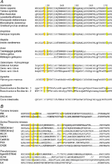
          Titin, the giant elastic ruler protein of striated muscle sarcomeres, contains a catalytic kinase domain related to a family of intrasterically regulated protein kinases. The most extensively studied member of this branch of the human kinome is the Ca 2+–calmodulin (CaM)-regulated myosin light-chain kinases (MLCK). However, not all kinases of the MLCK branch are functional MLCKs, and about half lack a CaM binding site in their C-terminal autoinhibitory tail (AI). A unifying feature is their association with the cytoskeleton, mostly via actin and myosin filaments. Titin kinase, similar to its invertebrate analogue twitchin kinase and likely other “MLCKs”, is not Ca 2+–calmodulin-activated. Recently, local protein unfolding of the C-terminal AI has emerged as a common mechanism in the activation of CaM kinases. Single-molecule data suggested that opening of the TK active site could also be achieved by mechanical unfolding of the AI. Mechanical modulation of catalytic activity might thus allow cytoskeletal signalling proteins to act as mechanosensors, creating feedback mechanisms between cytoskeletal tension and tension generation or cellular remodelling. Similar to other MLCK-like kinases like DRAK2 and DAPK1, TK is linked to protein turnover regulation via the autophagy/lysosomal system, suggesting the MLCK-like kinases have common functions beyond contraction regulation.

          Related collections

          Most cited references125

          • Record: found
          • Abstract: found
          • Article: not found

          The protein kinase complement of the human genome.

          G. Manning (2002)
          We have catalogued the protein kinase complement of the human genome (the "kinome") using public and proprietary genomic, complementary DNA, and expressed sequence tag (EST) sequences. This provides a starting point for comprehensive analysis of protein phosphorylation in normal and disease states, as well as a detailed view of the current state of human genome analysis through a focus on one large gene family. We identify 518 putative protein kinase genes, of which 71 have not previously been reported or described as kinases, and we extend or correct the protein sequences of 56 more kinases. New genes include members of well-studied families as well as previously unidentified families, some of which are conserved in model organisms. Classification and comparison with model organism kinomes identified orthologous groups and highlighted expansions specific to human and other lineages. We also identified 106 protein kinase pseudogenes. Chromosomal mapping revealed several small clusters of kinase genes and revealed that 244 kinases map to disease loci or cancer amplicons.
            Bookmark
            • Record: found
            • Abstract: found
            • Article: not found

            Reversible unfolding of individual titin immunoglobulin domains by AFM.

            Single-molecule atomic force microscopy (AFM) was used to investigate the mechanical properties of titin, the giant sarcomeric protein of striated muscle. Individual titin molecules were repeatedly stretched, and the applied force was recorded as a function of the elongation. At large extensions, the restoring force exhibited a sawtoothlike pattern, with a periodicity that varied between 25 and 28 nanometers. Measurements of recombinant titin immunoglobulin segments of two different lengths exhibited the same pattern and allowed attribution of the discontinuities to the unfolding of individual immunoglobulin domains. The forces required to unfold individual domains ranged from 150 to 300 piconewtons and depended on the pulling speed. Upon relaxation, refolding of immunoglobulin domains was observed.
              Bookmark
              • Record: found
              • Abstract: found
              • Article: not found

              A role for NBR1 in autophagosomal degradation of ubiquitinated substrates.

              Autophagy is a catabolic process where cytosolic cellular components are delivered to the lysosome for degradation. Recent studies have indicated the existence of specific receptors, such as p62, which link ubiquitinated targets to autophagosomal degradation pathways. Here we show that NBR1 (neighbor of BRCA1 gene 1) is an autophagy receptor containing LC3- and ubiquitin (Ub)-binding domains. NBR1 is recruited to Ub-positive protein aggregates and degraded by autophagy depending on an LC3-interacting region (LIR) and LC3 family modifiers. Although NBR1 and p62 interact and form oligomers, they can function independently, as shown by autophagosomal clearance of NBR1 in p62-deficient cells. NBR1 was localized to Ub-positive inclusions in patients with liver dysfunction, and depletion of NBR1 abolished the formation of Ub-positive p62 bodies upon puromycin treatment of cells. We propose that NBR1 and p62 act as receptors for selective autophagosomal degradation of ubiquitinated targets.
                Bookmark

                Author and article information

                Contributors
                +44-20-78486709 , +44-20-78486434 , mathias.gautel@kcl.ac.uk
                Journal
                Pflugers Arch
                Pflugers Archiv
                Springer-Verlag (Berlin/Heidelberg )
                0031-6768
                1432-2013
                18 March 2011
                18 March 2011
                July 2011
                : 462
                : 1
                : 119-134
                Affiliations
                King’s College London BHF Centre of Research Excellence, Cardiovascular Division and Randall Division for Cell and Molecular Biophysics, London, SE1 1UL UK
                Article
                946
                10.1007/s00424-011-0946-1
                3114093
                21416260
                537a9658-3dde-4733-9567-8c1fcf2fe201
                © The Author(s) 2011
                History
                : 4 February 2011
                : 15 February 2011
                : 18 February 2011
                Categories
                Invited Review
                Custom metadata
                © Springer-Verlag 2011

                Anatomy & Physiology
                obscurin,myomesin,p62/sqstm1,connectin,twitchin,murf,mechanobiology,autophagy,myosin light-chain kinase,nbr1,sarcomere,telethonin/tcap,mechanical strain sensor,titin

                Comments

                Comment on this article