14
views
0
recommends
+1 Recommend
0 collections
    0
    shares
      • Record: found
      • Abstract: found
      • Article: found
      Is Open Access

      Beyond the Classic VTA: Extended Amygdala Projections to DA-Striatal Paths in the Primate

      research-article

      Read this article at

      Bookmark
          There is no author summary for this article yet. Authors can add summaries to their articles on ScienceOpen to make them more accessible to a non-specialist audience.

          Abstract

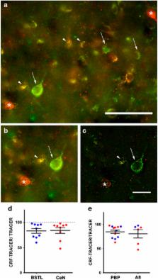
          The central extended amygdala (CEA) has been conceptualized as a ‘macrosystem’ that regulates various stress-induced behaviors. Consistent with this, the CEA highly expresses corticotropin-releasing factor (CRF), an important modulator of stress responses. Stress alters goal-directed responses associated with striatal paths, including maladaptive responses such as drug seeking, social withdrawal, and compulsive behavior. CEA inputs to the midbrain dopamine (DA) system are positioned to influence striatal functions through mesolimbic DA-striatal pathways. However, the structure of this amygdala-CEA-DA neuron path to the striatum has been poorly characterized in primates. In primates, we combined neuronal tracer injections into various arms of the circuit through specific DA subpopulations to assess: (1) whether the circuit connecting amygdala, CEA, and DA cells follows CEA intrinsic organization, or a more direct topography involving bed nucleus vs central nucleus divisions; (2) CRF content of the CEA-DA path; and (3) striatal subregions specifically involved in CEA-DA-striatal loops. We found that the amygdala-CEA-DA path follows macrostructural subdivisions, with the majority of input/outputs converging in the medial central nucleus, the sublenticular extended amygdala, and the posterior lateral bed nucleus of the stria terminalis. The proportion of CRF+ outputs is >50%, and mainly targets the A10 parabrachial pigmented nucleus (PBP) and A8 (retrorubal field, RRF) neuronal subpopulations, with additional inputs to the dorsal A9 neurons. CRF-enriched CEA-DA projections are positioned to influence outputs to the ‘limbic-associative’ striatum, which is distinct from striatal regions targeted by DA cells lacking CEA input. We conclude that the concept of the CEA is supported on connectional grounds, and that CEA termination over the PBP and RRF neuronal populations can influence striatal circuits involved in associative learning.

          Related collections

          Most cited references58

          • Record: found
          • Abstract: found
          • Article: not found

          Stress revisited: a critical evaluation of the stress concept.

          With the steadily increasing number of publications in the field of stress research it has become evident that the conventional usage of the stress concept bears considerable problems. The use of the term 'stress' to conditions ranging from even the mildest challenging stimulation to severely aversive conditions, is in our view inappropriate. Review of the literature reveals that the physiological 'stress' response to appetitive, rewarding stimuli that are often not considered to be stressors can be as large as the response to negative stimuli. Analysis of the physiological response during exercise supports the view that the magnitude of the neuroendocrine response reflects the metabolic and physiological demands required for behavioural activity. We propose that the term 'stress' should be restricted to conditions where an environmental demand exceeds the natural regulatory capacity of an organism, in particular situations that include unpredictability and uncontrollability. Physiologically, stress seems to be characterized by either the absence of an anticipatory response (unpredictable) or a reduced recovery (uncontrollable) of the neuroendocrine reaction. The consequences of this restricted definition for stress research and the interpretation of results in terms of the adaptive and/or maladaptive nature of the response are discussed. Copyright © 2011 Elsevier Ltd. All rights reserved.
            Bookmark
            • Record: found
            • Abstract: found
            • Article: not found

            Reward representations and reward-related learning in the human brain: insights from neuroimaging.

            This review outlines recent findings from human neuroimaging concerning the role of a highly interconnected network of brain areas including orbital and medial prefrontal cortex, amygdala, striatum and dopaminergic mid-brain in reward processing. Distinct reward-related functions can be attributed to different components of this network. Orbitofrontal cortex is involved in coding stimulus reward value and in concert with the amygdala and ventral striatum is implicated in representing predicted future reward. Such representations can be used to guide action selection for reward, a process that depends, at least in part, on orbital and medial prefrontal cortex as well as dorsal striatum.
              Bookmark
              • Record: found
              • Abstract: found
              • Article: not found

              Striatonigrostriatal pathways in primates form an ascending spiral from the shell to the dorsolateral striatum.

              Clinical manifestations in diseases affecting the dopamine system include deficits in emotional, cognitive, and motor function. Although the parallel organization of specific corticostriatal pathways is well documented, mechanisms by which dopamine might integrate information across different cortical/basal ganglia circuits are less well understood. We analyzed a collection of retrograde and anterograde tracing studies to understand how the striatonigrostriatal (SNS) subcircuit directs information flow between ventromedial (limbic), central (associative), and dorsolateral (motor) striatal regions. When viewed as a whole, the ventromedial striatum projects to a wide range of the dopamine cells and receives a relatively small dopamine input. In contrast, the dorsolateral striatum (DLS) receives input from a broad expanse of dopamine cells and has a confined input to the substantia nigra (SN). The central striatum (CS) receives input from and projects to a relatively wide range of the SN. The SNS projection from each striatal region contains three substantia nigra components: a dorsal group of nigrostriatal projecting cells, a central region containing both nigrostriatal projecting cells and its reciprocal striatonigral terminal fields, and a ventral region that receives a specific striatonigral projection but does not contain its reciprocal nigrostriatal projection. Examination of results from multiple tracing experiments simultaneously demonstrates an interface between different striatal regions via the midbrain dopamine cells that forms an ascending spiral between regions. The shell influences the core, the core influences the central striatum, and the central striatum influences the dorsolateral striatum. This anatomical arrangement creates a hierarchy of information flow and provides an anatomical basis for the limbic/cognitive/motor interface via the ventral midbrain.
                Bookmark

                Author and article information

                Journal
                Neuropsychopharmacology
                Neuropsychopharmacology
                Neuropsychopharmacology
                Nature Publishing Group
                0893-133X
                1740-634X
                July 2017
                21 February 2017
                22 March 2017
                1 July 2017
                : 42
                : 8
                : 1563-1576
                Affiliations
                [1 ]Department of Neuroscience, University of Rochester Medical Center , Rochester, NY, USA
                [2 ]Department of Psychiatry, University of Rochester Medical Center , Rochester, NY, USA
                [3 ]Department of Neuroscience, University of Pennsylvania , Philadelphia, PA, USA
                [4 ]Jacobs School of Medicine and Biomedical Sciences, State University of New York at Buffalo , Buffalo, NY, USA
                [5 ]Boston University School of Medicine , Boston, MA, USA
                Author notes
                [* ]Departments of Neuroscience and Psychiatry, University of Rochester Medical Center , 601 Elmwood Avenue, Box 601, Rochester, NY 14642, USA. Tel: +1 585 273 2028, Fax: +1 585 754 5336, E-mail: Julie_Fudge@ 123456urmc.rochester.edu
                Article
                npp201738
                10.1038/npp.2017.38
                5518904
                28220796
                538a295b-7ab4-469c-ac1d-64cd52a9cd0f
                Copyright © 2017 The Author(s)

                This work is licensed under a Creative Commons Attribution-NonCommercial-NoDerivs 4.0 International License. The images or other third party material in this article are included in the article’s Creative Commons license, unless indicated otherwise in the credit line; if the material is not included under the Creative Commons license, users will need to obtain permission from the license holder to reproduce the material. To view a copy of this license, visit http://creativecommons.org/licenses/by-nc-nd/4.0/

                History
                : 17 June 2016
                : 08 February 2017
                : 12 February 2017
                Categories
                Original Article

                Pharmacology & Pharmaceutical medicine
                Pharmacology & Pharmaceutical medicine

                Comments

                Comment on this article