21
views
0
recommends
+1 Recommend
0 collections
    0
    shares
      • Record: found
      • Abstract: found
      • Article: not found

      The influence of low-grade glioma on resting state oscillatory brain activity: a magnetoencephalography study

      research-article

      Read this article at

      ScienceOpenPublisherPMC
      Bookmark
          There is no author summary for this article yet. Authors can add summaries to their articles on ScienceOpen to make them more accessible to a non-specialist audience.

          Abstract

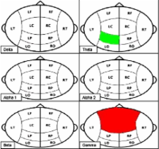
          Purpose In the present MEG-study, power spectral analysis of oscillatory brain activity was used to compare resting state brain activity in both low-grade glioma (LGG) patients and healthy controls. We hypothesized that LGG patients show local as well as diffuse slowing of resting state brain activity compared to healthy controls and that particularly global slowing correlates with neurocognitive dysfunction. Patient and methods Resting state MEG recordings were obtained from 17 LGG patients and 17 age-, sex-, and education-matched healthy controls. Relative spectral power was calculated in the delta, theta, upper and lower alpha, beta, and gamma frequency band. A battery of standardized neurocognitive tests measuring 6 neurocognitive domains was administered. Results LGG patients showed a slowing of the resting state brain activity when compared to healthy controls. Decrease in relative power was mainly found in the gamma frequency band in the bilateral frontocentral MEG regions, whereas an increase in relative power was found in the theta frequency band in the left parietal region. An increase of the relative power in the theta and lower alpha band correlated with impaired executive functioning, information processing, and working memory. Conclusion LGG patients are characterized by global slowing of their resting state brain activity and this slowing phenomenon correlates with the observed neurocognitive deficits.

          Related collections

          Most cited references37

          • Record: found
          • Abstract: found
          • Article: not found

          Neural synchrony in brain disorders: relevance for cognitive dysfunctions and pathophysiology.

          Following the discovery of context-dependent synchronization of oscillatory neuronal responses in the visual system, novel methods of time series analysis have been developed for the examination of task- and performance-related oscillatory activity and its synchronization. Studies employing these advanced techniques revealed that synchronization of oscillatory responses in the beta- and gamma-band is involved in a variety of cognitive functions, such as perceptual grouping, attention-dependent stimulus selection, routing of signals across distributed cortical networks, sensory-motor integration, working memory, and perceptual awareness. Here, we review evidence that certain brain disorders, such as schizophrenia, epilepsy, autism, Alzheimer's disease, and Parkinson's are associated with abnormal neural synchronization. The data suggest close correlations between abnormalities in neuronal synchronization and cognitive dysfunctions, emphasizing the importance of temporal coordination. Thus, focused search for abnormalities in temporal patterning may be of considerable clinical relevance.
            Bookmark
            • Record: found
            • Abstract: found
            • Article: not found

            Cognitive deficits in adult patients with brain tumours.

            Cognitive function, with survival and response on brain imaging, is increasingly regarded as an important outcome measure in patients with brain tumours. This measure provides us with information on a patient's clinical situation and adverse treatment effects. Radiotherapy has been regarded as the main cause of cognitive decline in these patients, because children with brain tumours can develop intellectual deterioration caused by radiotherapy. In long-term surviving patients, radiotherapy may indeed lead to cognitive deficits, or even dementia. Recent studies, however, have made clear that focal radiotherapy in patients with glioma is not the main reason for cognitive deficits. The tumour itself and other medical treatments contribute largely to the cognitive deficits. Cognitive function is now also recognised as an independent prognostic factor in the survival of glioma patients. Additionally, cognitive deterioration can be the first indicator of progressive disease after treatment.
              Bookmark
              • Record: found
              • Abstract: found
              • Article: not found

              Slowing of oscillatory brain activity is a stable characteristic of Parkinson's disease without dementia.

              Extensive changes in resting-state oscillatory brain activity have recently been demonstrated using magnetoencephalography (MEG) in moderately advanced, non-demented Parkinson's disease patients relative to age-matched controls. The aim of the present study was to determine the onset and evolution of these changes over the disease course and their relationship with clinical parameters. In addition, we evaluated the effects of dopaminomimetics on resting-state oscillatory brain activity in levodopa-treated patients. MEG background oscillatory activity was studied in a group of 70 Parkinson's disease patients with varying disease duration and severity (including 18 de novo patients) as well as in 21 controls that were age-matched to the de novo patients. Whole head 151-channel MEG recordings were obtained in an eyes-closed resting-state condition. Levodopa-treated patients (N = 37) were examined both in a practically defined 'OFF' as well as in the 'ON' state. Relative spectral power was calculated for delta, theta, low alpha, high alpha, beta and gamma frequency bands and averaged for 10 cortical regions of interest (ROIs). Additionally, extensive clinical and neuropsychological testing was performed in all subjects. De novo Parkinson's disease patients showed widespread slowing of background MEG activity relative to controls. Changes included a widespread increase in theta and low alpha power, as well as a loss of beta power over all but the frontal ROIs and a loss of gamma power over all but the right occipital ROI. Neuropsychological assessment revealed abnormal perseveration in de novo patients, which was associated with increased low alpha power in centroparietal ROIs. In the whole group of Parkinson's disease patients, longer disease duration was associated with reduced low alpha power in the right temporal and right occipital ROI, but not with any other spectral power measure. No association was found between spectral power and disease stage, disease severity or dose of dopaminomimetics. In patients on levodopa therapy, a change from the 'OFF' to the 'ON' state was associated with decreases in right frontal theta, left occipital beta and left temporal gamma power and an increase in right parietal gamma power. Widespread slowing of oscillatory brain activity is a characteristic of non-demented Parkinson's disease patients from the earliest clinical stages onwards that is (largely) independent of disease duration, stage and severity and hardly influenced by dopaminomimetic treatment. Some early cognitive deficits in Parkinson's disease appear to be associated with increased low alpha power. We postulate a role for hypofunctional non-dopaminergic ascending neurotransmitter systems in spectral power changes in non-demented Parkinson's disease patients.
                Bookmark

                Author and article information

                Contributors
                +31-20-4440183 , +31-20-4440715 , i.bosma@vumc.nl
                Journal
                J Neurooncol
                Journal of Neuro-Oncology
                Springer US (Boston )
                0167-594X
                1573-7373
                8 February 2008
                May 2008
                : 88
                : 1
                : 77-85
                Affiliations
                [1 ]Department of Neurology, VU University Medical Center, De Boelelaan 1117, Amsterdam, 1081 HV The Netherlands
                [2 ]Department of Clinical Neurophysiology, VU University Medical Center, Amsterdam, The Netherlands
                [3 ]Department of Medical Psychology, VU University Medical Center, Amsterdam, The Netherlands
                [4 ]Department of Clinical Neurophysiology and Epileptology, Centre Hospitalier Universitaire Timone, Marseille, France
                [5 ]Magnetoencephalography Center, VU University Medical Center, Amsterdam, The Netherlands
                [6 ]Department of Neurology, Academic Medical Center, Amsterdam, The Netherlands
                Article
                9535
                10.1007/s11060-008-9535-3
                2295256
                18259691
                54ccaad8-86db-4725-bdbd-f462d34f71a8
                © The Author(s) 2008
                History
                : 27 July 2007
                : 21 January 2008
                Categories
                Clinical-Patient Studies
                Custom metadata
                © Springer Science+Business Media, LLC. 2008

                Oncology & Radiotherapy
                meg,low-grade glioma,cognition,power analysis
                Oncology & Radiotherapy
                meg, low-grade glioma, cognition, power analysis

                Comments

                Comment on this article