52
views
0
recommends
+1 Recommend
0 collections
    0
    shares
      • Record: found
      • Abstract: found
      • Article: found
      Is Open Access

      Identification of a Common Non-Apoptotic Cell Death Mechanism in Hereditary Retinal Degeneration

      research-article

      Read this article at

      Bookmark
          There is no author summary for this article yet. Authors can add summaries to their articles on ScienceOpen to make them more accessible to a non-specialist audience.

          Abstract

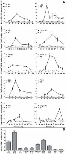
          Cell death in neurodegenerative diseases is often thought to be governed by apoptosis; however, an increasing body of evidence suggests the involvement of alternative cell death mechanisms in neuronal degeneration. We studied retinal neurodegeneration using 10 different animal models, covering all major groups of hereditary human blindness ( rd1, rd2, rd10, Cngb1 KO, Rho KO, S334ter, P23H, Cnga3 KO, cpfl1, Rpe65 KO), by investigating metabolic processes relevant for different forms of cell death. We show that apoptosis plays only a minor role in the inherited forms of retinal neurodegeneration studied, where instead, a non-apoptotic degenerative mechanism common to all mutants is of major importance. Hallmark features of this pathway are activation of histone deacetylase, poly-ADP-ribose-polymerase, and calpain, as well as accumulation of cyclic guanosine monophosphate and poly-ADP-ribose. Our work thus demonstrates the prevalence of alternative cell death mechanisms in inherited retinal degeneration and provides a rational basis for the design of mutation-independent treatments.

          Related collections

          Most cited references52

          • Record: found
          • Abstract: found
          • Article: not found

          Identification of programmed cell death in situ via specific labeling of nuclear DNA fragmentation

          Programmed cell death (PCD) plays a key role in developmental biology and in maintenance of the steady state in continuously renewing tissues. Currently, its existence is inferred mainly from gel electrophoresis of a pooled DNA extract as PCD was shown to be associated with DNA fragmentation. Based on this observation, we describe here the development of a method for the in situ visualization of PCD at the single-cell level, while preserving tissue architecture. Conventional histological sections, pretreated with protease, were nick end labeled with biotinylated poly dU, introduced by terminal deoxy- transferase, and then stained using avidin-conjugated peroxidase. The reaction is specific, only nuclei located at positions where PCD is expected are stained. The initial screening includes: small and large intestine, epidermis, lymphoid tissues, ovary, and other organs. A detailed analysis revealed that the process is initiated at the nuclear periphery, it is relatively short (1-3 h from initiation to cell elimination) and that PCD appears in tissues in clusters. The extent of tissue-PCD revealed by this method is considerably greater than apoptosis detected by nuclear morphology, and thus opens the way for a variety of studies.
            Bookmark
            • Record: found
            • Abstract: found
            • Article: not found

            Cleavage of poly(ADP-ribose) polymerase by a proteinase with properties like ICE.

            Recent studies suggest that proteases of the interleukin 1-beta-converting enzyme (ICE)/ced-3 family are involved in initiating the active phase of apoptosis. Here we identify a novel protease resembling ICE (prICE) that is active in a cell-free system that reproduces the morphological and biochemical events of apoptosis. prICE cleaves the nuclear enzyme poly(ADP-ribose) polymerase (PARP) at a tetrapeptide sequence identical to one of two ICE sites in pro-interleukin-1-beta. However, prICE does not cleave purified pro-interleukin-1-beta, and purified ICE does not cleave PARP, indicating that the two activities are distinct. Inhibition of prICE abolishes all manifestations of apoptosis in the extracts including morphological changes, cleavage of PARP and production of an oligonucleosomal ladder. These studies suggest that prICE might be pivotal in initiating the active phase of apoptosis in vitro and in intact cells.
              Bookmark
              • Record: found
              • Abstract: found
              • Article: not found

              Poly(ADP-ribose) (PAR) binding to apoptosis-inducing factor is critical for PAR polymerase-1-dependent cell death (parthanatos).

              The mitochondrial protein apoptosis-inducing factor (AIF) plays a pivotal role in poly(ADP-ribose) polymerase-1 (PARP-1)-mediated cell death (parthanatos), during which it is released from the mitochondria and translocates to the nucleus. We show that AIF is a high-affinity poly(ADP-ribose) (PAR)-binding protein and that PAR binding to AIF is required for parthanatos both in vitro and in vivo. AIF bound PAR at a site distinct from AIF's DNA binding site, and this interaction triggered AIF release from the cytosolic side of the mitochondrial outer membrane. Mutation of the PAR binding site in AIF did not affect its NADH (reduced form of nicotinamide adenine dinucleotide) oxidase activity, its ability to bind FAD (flavin adenine dinucleotide) or DNA, or its ability to induce nuclear condensation. However, this AIF mutant was not released from mitochondria and did not translocate to the nucleus or mediate cell death after PARP-1 activation. These results suggest a mechanism for PARP-1 to initiate AIF-mediated cell death and indicate that AIF's bioenergetic cell survival-promoting functions are separate from its effects as a mitochondrially derived death effector. Interference with the PAR-AIF interaction or PAR signaling may provide notable opportunities for preventing cell death after activation of PARP-1.
                Bookmark

                Author and article information

                Contributors
                Role: Editor
                Journal
                PLoS One
                PLoS ONE
                plos
                plosone
                PLoS ONE
                Public Library of Science (San Francisco, USA )
                1932-6203
                2014
                13 November 2014
                : 9
                : 11
                : e112142
                Affiliations
                [1 ]Institute for Ophthalmic Research, University of Tuebingen, Tuebingen, Germany
                [2 ]Department of Neurobiology, University of Oldenburg, Oldenburg, Germany
                [3 ]Center for Integrated Protein Science Munich and Department of Pharmacy - Center for Drug Research, Ludwig-Maximilians-University Munich, Munich, Germany
                [4 ]Division of Ophthalmology, Department of Clinical Sciences, University of Lund, Lund, Sweden
                [5 ]Institute for Research in Ophthalmology, Sion, Switzerland
                [6 ]Centre for Integrative Neuroscience, University of Tuebingen, Tuebingen, Germany
                Eye Hospital, Charité, Germany
                Author notes

                Competing Interests: The authors declare that they have no conflict of interest.

                Conceived and designed the experiments: BAG FPD. Performed the experiments: BAG DT AS KK PF FPD. Analyzed the data: BAG DT AS KK SC PF KD EZ TE MU FPD. Contributed reagents/materials/analysis tools: BAG SM SK FK SC UJB KD MB TE PE FPD. Wrote the paper: BAG DT FPD.

                Article
                PONE-D-14-25834
                10.1371/journal.pone.0112142
                4230983
                25392995
                5664bfe0-562f-4a40-818c-02bb532d8490
                Copyright @ 2014

                This is an open-access article distributed under the terms of the Creative Commons Attribution License, which permits unrestricted use, distribution, and reproduction in any medium, provided the original author and source are credited.

                History
                : 12 June 2014
                : 13 October 2014
                Page count
                Pages: 11
                Funding
                This work was supported by the Kerstan Foundation, Deutsche Forschungsgemeinschaft [DFG PA1751/1-1, 4-1], Alcon Research Institute, European Commission [DRUGSFORD: HEALTH-F2-2012-304963; PANOPTES: NMP4-SL-2010-246180], German Ministry of Education and Research [BMBF HOPE2 - FKZ 01GM1108A], Centre for Integrative Neuroscience [CIN pool project 2009-20], Kronprinsessan Margaretas Arbetsnämnd (KMA), Stiftelsen Olle Engkvist Byggmästare, The Swedish Research Council 2009-3855, and Stiftelsen för Synskadade i f.d. Malmöhus län. The funders had no role in study design, data collection and analysis, decision to publish, or preparation of the manuscript.
                Categories
                Research Article
                Biology and Life Sciences
                Cell Biology
                Cell Processes
                Cell Death
                Apoptosis
                Necrotic Cell Death
                Neuronal Death
                Medicine and Health Sciences
                Ophthalmology
                Retinal Disorders
                Retinal Degeneration
                Inherited Eye Disorders
                Custom metadata
                The authors confirm that all data underlying the findings are fully available without restriction. All relevant data are within the paper and its Supporting Information files.

                Uncategorized
                Uncategorized

                Comments

                Comment on this article