- Record: found
- Abstract: found
- Article: found
A novel mapping technique to detect non–pulmonary vein triggers: A case report of self-reference mapping technique
case-report
06 November 2017
Read this article at
There is no author summary for this article yet. Authors can add summaries to their articles on ScienceOpen to make them more accessible to a non-specialist audience.
Abstract
Key Teaching Points
•
Self-reference mapping with the PentaRay NAV catheter is useful to detect non–pulmonary
vein triggers.
•
This new mapping technique uses the previous earliest electrode site as the reference
and does not require other reference catheters.
•
The earliest site should be distinguished from other later activated sites. This map,
the map to identify the earliest site, size is considered to be within the range of
the PentaRay catheter size.
Introduction
Catheter ablation is an effective therapeutic strategy for atrial fibrillation (AF).
Pulmonary vein (PV) isolation is the cornerstone of catheter ablation for AF. After
all PVs were isolated, ablation of non-PV triggers should be attempted. Successful
elimination of all the possible AF triggers is considered a better outcome.1, 2 Mapping
of non-PV triggers is usually performed using 3-dimensional anatomic mapping; however,
precise mapping of non-PV triggers is sometimes difficult. The electrogram obtained
using the reference catheter is not suitable to use as the reference, because of poor
stability of the catheter, ventricular electrogram overlap, or dull potential after
cardioversion. A new technique, self-reference mapping, does not require other reference
catheters, because it uses the previous earliest activation site recorded with the
PentaRay NAV (PEN) catheter (Biosense Webster Inc., Diamond Bar, CA) as the reference
(Figure 1).
Figure 1
Theoretical illustration of self-reference mapping. A: A non–pulmonary vein trigger
is considered to conduct centrifugally. The earliest activation site of the PentaRay
catheter, which is located far from the trigger, should be one of the outer electrodes
(1-2, 5-6, 9-10, 13-14, 17-18). B: The PentaRay catheter is moved to the earlier activation
site and placed including the previous earliest site, which is used as the reference
for the next trigger mapping method. C: After several mapping iterations, one of the
inner electrodes (3-4, 7-8, 11-12, 15-16, 19-20) of the PentaRay catheter can record
the earliest activation site. It means that activation of all the outer electrodes
is late. D: When the earliest activation tags are concentrated in a small area, the
area is the origin of the trigger.
Case report
A 75-year-old man with a 2-month history of persistent AF and transient ischemic attack
was referred to our institution for catheter ablation. Catheter ablation was performed
using an open-irrigated contact-force catheter (ThermoCool SmartTouch SF, Biosense
Webster) with an electroanatomic mapping system (CARTO 3, Biosense Webster). After
circumferential bilateral PV isolation and superior vena cava isolation, continuous
isoproterenol infusion (2 μg/min) and adenosine triphosphate (ATP) rapid injection
(30 mg) were performed to provoke non-PV trigger.
3
AF was induced by ATP administration, and spontaneous AF initiation was reproducibly
observed by the postcardioversion AF trigger.
Self-reference mapping was performed as follows. As the first step, PEN was located
at the low left atrial septum side because the earliest activation site of the non-PV
trigger except PEN was the proximal coronary sinus. The earliest activation site of
the first non-PV trigger was PEN 17-18. The red tag was placed at PEN 17-18 (Figure 2A).
The ablation catheter was located in the right atrium at the opposite site of PEN
(Figure 2A). Activation of the ablation catheter was later than that of PEN, and following
mapping iterations were performed in the left atrium.
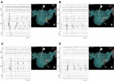
Figure 2
Steps of self-reference mapping. A: PEN was located at the low left atrial septum
side. The earliest activation site of the non-PV trigger was PEN 17-18. The red tag
was placed at PEN 17-18. B: PEN was moved to a higher position including the previous
red tag. The earliest activation sites of the next non-PV trigger were PEN 13-14 and
PEN 15-16. The yellow tag was placed at PEN 15-16, and the red tag was placed at PEN
13-14. C: PEN was moved to the anterior position. The earliest activation site of
the next non-PV trigger was PEN 15-16. The yellow tag was placed at PEN 15-16. The
earliest tags obtained by inner electrodes were concentrated in a small area. The
area was supposed to be the origin of the non-PV trigger. D: PEN was moved more anterior,
including the previous earliest yellow tag to confirm the accuracy of the map. The
earliest activation site was PEN 9-10, which was the same as the previous earliest
activation site. ABL = ablation catheter; CS = coronary sinus; d = distal; p = proximal;
PEN = PentaRay; PV = pulmonary vein; RA = right atrium; SVC = superior vena cava.
PEN was moved to a higher position including the previous red tag. The earliest activation
sites of the next non-PV trigger were PEN 13-14 and PEN 15-16. The yellow tag was
placed at PEN 15-16, and the red tag was placed at PEN 13-14 (Figure 2B). PEN was
moved to the anterior position. The earliest activation site of the next non-PV trigger
was PEN 15-16. The yellow tag was placed at PEN 15-16. The earliest tags obtained
by inner electrodes of PEN were concentrated in a small area. The area was supposed
to be the origin of the non-PV trigger (Figure 2C). PEN was moved more anterior, including
the previous earliest yellow tag to confirm the accuracy of the map. The earliest
activation site of the next non-PV trigger was PEN 9-10, which was the same place
as the previous earliest activation site (Figure 2D). During self-reference mapping,
the operator has to pay close attention to the intracardiac activation sequence of
beat triggering AF as well as the coupling interval in order to distinguish a true
spontaneous trigger from catheter-induced ectopy.
Ablation was performed during AF to cover the yellow tag sites maintaining contact
force at least 10 g (25 W, 25 seconds at each point). The end point of ablation was
noninducibility of AF after cardioversion. After 1 series of ablation (7 points),
AF was never induced by ATP administration (ATP administrations were performed 7 times
and AF was never induced) during continuous isoproterenol infusion.
Discussion
The main goal of catheter ablation for AF is elimination of all the possible triggers
from both PV and non-PV, which typically arise from a discrete anatomical structure.
4
Because a non-PV trigger conducts centrifugally, the earliest site should be distinguished
from other later activated sites. This map, the map to identify the earliest site,
size is considered to be within the range of the PEN catheter size. In this article,
we proposed a new self-reference mapping technique to detect non-PV triggers.
Our technique is similar to the previously reported vector mapping technique, which
is performed to characterize atrial tachycardia.
5
Vector mapping is performed to identify the earliest activation site comparing PentaRay
NAV catheter electrogram with the P wave and fixed catheter within coronary sinus.
The concept of searching the core of the centrifugal activation pattern by focal atrial
tachycardia or non-PV triggers is the same. Vector mapping is performed with regard
to localized reentry and focal atrial tachycardia. Our technique is specialized for
non-PV trigger mapping and does not require the reference catheter. Moreover, a recently
developed 3-dimensional anatomic mapping technique made it possible to obtain a tag
by a multielectrode catheter. These 2 advantages led us to make a precise high-resolution
map in a small area easily.
There are some limitations of this mapping method. This mapping method needs reproducible
AF initiation from the same site. When multiple triggers were observed, we tried to
map 1 by 1, assessing the intracardiac activation sequence. We do not have experience
using this method in cases post excessive atrial ablation, such as CFAE or step wise
substrate ablation, because we do not perform these kinds of ablation methods at our
institution. In these cases, conduction of the atrial myocardium might be significantly
affected by excessive ablation and the impaired electrogram postablation might be
not suitable for assessing the conduction pattern. A study is needed to elucidate
the accuracy, success rate, and safety of the self-reference mapping method.
Conclusion
This case demonstrated the usefulness of self-reference mapping to detect a non-PV
trigger induced by isoproterenol and adenosine triphosphate.
Related collections
Most cited references4
- Record: found
- Abstract: found
- Article: not found
Trigger-based mechanism of the persistence of atrial fibrillation and its impact on the efficacy of catheter ablation.
Koichi Inoue, Toshiya Kurotobi, Ryusuke Kimura … (2012)
- Record: found
- Abstract: found
- Article: not found
Impact of Non-Pulmonary Vein Foci on the Outcome of the Second Session of Catheter Ablation for Paroxysmal Atrial Fibrillation.
- Record: found
- Abstract: found
- Article: not found