3
views
0
recommends
+1 Recommend
0 collections
    0
    shares
      • Record: found
      • Abstract: found
      • Article: found
      Is Open Access

      High detection rate in [ 18F]PSMA-1007 PET: interim results focusing on biochemical recurrence in prostate cancer patients

      brief-report

      Read this article at

      Bookmark
          There is no author summary for this article yet. Authors can add summaries to their articles on ScienceOpen to make them more accessible to a non-specialist audience.

          Abstract

          Objective

          18F-labeled prostate-specific membrane antigen (PSMA) ligand, [ 18F]PSMA-1007, has the benefit of a higher synthetic yield and minimal excretion in the urine. High detection efficacy was reported in biochemical recurrence (BCR) of prostate cancer after radical prostatectomy. Thus, we evaluated the preliminary diagnostic utility of [ 18F]PSMA-1007 PET in patients with prostate cancer, focusing on the BCR which is not detected on conventional imaging.

          Methods

          We enrolled a total of 28 patients (age 51–79 years) with BCR of prostate cancer. BCR was defined as a continuous increase in PSA after radical prostatectomy or radiation therapy without any apparent recurrent lesions on conventional diagnostic imaging (CT and bone scintigraphy). PSMA-PET scanning was performed approximately 60 min after intravenous injection of [ 18F]PSMA-1007 (259 ± 37 MBq). PSMA-PET images were evaluated for lesion detection as well as its relation to PSA values and location.

          Results

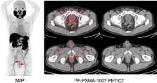
          Abnormal uptake, which was suspected to be recurrence or metastasis, was detected in 92.9% (26/28) of patients with BCR. The SUVmax was 8.4 ± 6.4 in local recurrence, 11.5 ± 11.8 in pelvic lymph nodes (LN), and 4.1 ± 1.6 in bone metastasis. The detection rates were 66.7% in the PSA group-1 (0.1–0.5 ng/mL), 85.7% in the PSA group-2 (0.5–1.0 ng/mL), and 100% in the PSA group-3 (above 1.0 ng/mL). Among the PET-positive BCR patients ( n = 26), local recurrence was detected in 57.7% (15/26), pelvic LN in 42.3% (11/26), and bone metastasis in 15.4% (4/26). In 53% (8/15) of BCR patients who were suspected of local recurrence, focal uptake was detected adjacent to the bladder on [ 18F]PSMA-1007 PET. This suggested the significant advantage of having minimal physiological urine excretion.

          Conclusions

          [ 18F]PSMA-1007 PET showed a high detection rate in recurrent and metastatic lesions. In patients with BCR, its high detection led to suitable treatment strategies, such as salvage radiation therapy or surgical removal of recurrent lymph nodes.

          Trial registration

          (UMIN Clinical Trials Registry) UMIN000037697.

          Related collections

          Most cited references13

          • Record: found
          • Abstract: found
          • Article: found
          Is Open Access

          F-18 labelled PSMA-1007: biodistribution, radiation dosimetry and histopathological validation of tumor lesions in prostate cancer patients

          Purpose The prostate-specific membrane antigen (PSMA) targeted positron-emitting-tomography (PET) tracer 68Ga-PSMA-11 shows great promise in the detection of prostate cancer. However, 68Ga has several shortcomings as a radiolabel including short half-life and non-ideal energies, and this has motivated consideration of 18F-labelled analogs. 18F-PSMA-1007 was selected among several 18F-PSMA-ligand candidate compounds because it demonstrated high labelling yields, outstanding tumor uptake and fast, non-urinary background clearance. Here, we describe the properties of 18F-PSMA-1007 in human volunteers and patients. Methods Radiation dosimetry of 18F-PSMA-1007 was determined in three healthy volunteers who underwent whole-body PET-scans and concomitant blood and urine sampling. Following this, ten patients with high-risk prostate cancer underwent 18F-PSMA-1007 PET/CT (1 h and 3 h p.i.) and normal organ biodistribution and tumor uptakes were examined. Eight patients underwent prostatectomy with extended pelvic lymphadenectomy. Uptake in intra-prostatic lesions and lymph node metastases were correlated with final histopathology, including PSMA immunostaining. Results With an effective dose of approximately 4.4–5.5 mSv per 200–250 MBq examination, 18F-PSMA-1007 behaves similar to other PSMA-PET agents as well as to other 18F-labelled PET-tracers. In comparison to other PSMA-targeting PET-tracers, 18F-PSMA-1007 has reduced urinary clearance enabling excellent assessment of the prostate. Similar to 18F-DCFPyL and with slightly slower clearance kinetics than PSMA-11, favorable tumor-to-background ratios are observed 2–3 h after injection. In eight patients, diagnostic findings were successfully validated by histopathology. 18F-PSMA-1007 PET/CT detected 18 of 19 lymph node metastases in the pelvis, including nodes as small as 1 mm in diameter. Conclusion 18F-PSMA-1007 performs at least comparably to 68Ga-PSMA-11, but its longer half-life combined with its superior energy characteristics and non-urinary excretion overcomes some practical limitations of 68Ga-labelled PSMA-targeted tracers. Electronic supplementary material The online version of this article (doi:10.1007/s00259-016-3573-4) contains supplementary material, which is available to authorized users.
            Bookmark
            • Record: found
            • Abstract: found
            • Article: found
            Is Open Access

            Detection Efficacy of 18 F-PSMA-1007 PET/CT in 251 Patients with Biochemical Recurrence of Prostate Cancer After Radical Prostatectomy

            Prostate-specific membrane antigen (PSMA)–targeted PET imaging recently emerged as a new method for the staging and restaging of prostate cancer. Most published studies investigated the diagnostic potential of 68Ga-labeled PSMA agents that are excreted renally. 18F-PSMA-1007 is a novel PSMA ligand that has excellent preclinical characteristics and that is only minimally excreted by the urinary tract, a potential advantage for pelvic imaging. The aim of this study was to investigate the diagnostic efficacy of 18F-PSMA-1007 for biochemical recurrence (BCR) after radical prostatectomy. Methods: From 3 academic centers, 251 patients with BCR after radical prostatectomy were evaluated in a retrospective analysis. Patients who had received second-line androgen deprivation therapy (ADT) or chemotherapy were excluded, but prior first-line ADT exposure was allowed. The median prostate-specific antigen (PSA) level was 1.2 ng/mL (range, 0.2–228 ng/mL). All patients underwent PSMA PET/CT at 92 ± 26 min after injection of 301 ± 46 MBq of 18F-PSMA-1007. The rate of detection of presumed recurrence sites was correlated with the PSA level and original primary Gleason score. A comparison to a subset of patients treated previously with ADT was undertaken. Results: Of the 251 patients, 204 (81.3%) had evidence of recurrence on 18F-PSMA-1007 PET/CT. The detection rates were 94.0% (79/84), 90.9% (50/55), 74.5% (35/47), and 61.5% (40/65) for PSA levels of greater than or equal to 2, 1 to less than 2, 0.5 to less than 1, and 0.2 to less than 0.5 ng/mL, respectively. 18F-PSMA-1007 PET/CT revealed local recurrence in 24.7% of patients (n = 62). Lymph node metastases were present in the pelvis in 40.6% of patients (n = 102), in the retroperitoneum in 19.5% of patients (n = 49), and in supradiaphragmatic locations in 12.0% of patients (n = 30). Bone and visceral metastases were detected in 40.2% of patients (n = 101) and in 3.6% of patients (n = 9), respectively. In tumors with higher Gleason scores (≤7 vs. ≥8), detection efficacy trended higher (76.3% vs. 86.7%) but was not statistically significant (P = 0.32). However, detection efficacy was higher in patients who had received ADT (91.7% vs. 78.0%) within 6 mo before imaging (P = 0.0179). Conclusion: 18F-PSMA-1007 PET/CT offers high detection rates for BCR after radical prostatectomy that are comparable to or better than those published for 68Ga-labeled PSMA ligands.
              Bookmark
              • Record: found
              • Abstract: found
              • Article: not found

              Diagnostic performance of 68Ga-PSMA-11 (HBED-CC) PET/CT in patients with recurrent prostate cancer: evaluation in 1007 patients

              Purpose Since the clinical introduction of 68Ga-PSMA-11 PET/CT, this imaging method has rapidly spread and is now regarded as a significant step forward in the diagnosis of recurrent prostate cancer (PCa). The aim of this study was to analyse the influence of several variables with possible influence on PSMA ligand uptake in a large cohort. Methods We performed a retrospective analysis of 1007 consecutive patients who were scanned with 68Ga-PSMA-11 PET/CT (1 h after injection) from January 2014 to January 2017 to detect recurrent disease. Patients with untreated primary PCa or patients referred for PSMA radioligand therapy were excluded. The possible effects of different variables including PSA level and PSA doubling time (PSADT), PSA velocity (PSAVel), Gleason score (GSC, including separate analysis of GSC 7a and 7b), ongoing androgen deprivation therapy (ADT), patient age and amount of injected activity were evaluated. Results In 79.5% of patients at least one lesion with characteristics suggestive of recurrent PCa was detected. A pathological (positive) PET/CT scan was associated with PSA level and ADT. GSC, amount of injected activity, patient age, PSADT and PSAVel were not associated with a positive PET/CT scan in multivariate analysis. Conclusion 68Ga-PSMA-11 PET/CT detects tumour lesions in a high percentage of patients with recurrent PCa. Tumour detection is clearly associated with PSA level and ADT. Only a tendency for an association without statistical significance was found between higher GSC and a higher probability of a pathological PET/CT scan. No associations were found between a pathological 68Ga-PSMA-11 PET/CT scan and patient age, amount of injected activity, PSADT or PSAVel. Electronic supplementary material The online version of this article (doi:10.1007/s00259-017-3711-7) contains supplementary material, which is available to authorized users.
                Bookmark

                Author and article information

                Contributors
                watabe@tracer.med.osaka-u.ac.jp
                Journal
                Ann Nucl Med
                Ann Nucl Med
                Annals of Nuclear Medicine
                Springer Singapore (Singapore )
                0914-7187
                1864-6433
                4 March 2021
                4 March 2021
                2021
                : 35
                : 4
                : 523-528
                Affiliations
                [1 ]GRID grid.136593.b, ISNI 0000 0004 0373 3971, Department of Nuclear Medicine and Tracer Kinetics, Graduate School of Medicine, , Osaka University, ; 2-2 Yamadaoka, Suita, Osaka 565-0871 Japan
                [2 ]GRID grid.136593.b, ISNI 0000 0004 0373 3971, Institute for Radiation Sciences, , Osaka University, ; Osaka, Japan
                [3 ]GRID grid.136593.b, ISNI 0000 0004 0373 3971, Department of Urology, Graduate School of Medicine, , Osaka University, ; Osaka, Japan
                [4 ]GRID grid.412398.5, ISNI 0000 0004 0403 4283, Department of Radiology, , Osaka University Hospital, ; Osaka, Japan
                [5 ]GRID grid.136593.b, ISNI 0000 0004 0373 3971, Department of Molecular Imaging in Medicine, Graduate School of Medicine, , Osaka University, ; 2-2 Yamadaoka, Suita, Osaka 565-0871 Japan
                [6 ]GRID grid.5253.1, ISNI 0000 0001 0328 4908, Department of Nuclear Medicine, , University Hospital Heidelberg, ; Heidelberg, Germany
                [7 ]GRID grid.265073.5, ISNI 0000 0001 1014 9130, Department of Diagnostic Radiology, , Medical Hospital, Tokyo Medical and Dental University, ; Tokyo, Japan
                [8 ]GRID grid.14778.3d, ISNI 0000 0000 8922 7789, Department of Nuclear Medicine, , University Hospital Düsseldorf, ; Düsseldorf, Germany
                Author information
                http://orcid.org/0000-0001-8658-2395
                Article
                1602
                10.1007/s12149-021-01602-x
                7981319
                33661475
                592f2e39-f5c8-45d2-960a-2ecc680d03f3
                © The Author(s) 2021

                Open AccessThis article is licensed under a Creative Commons Attribution 4.0 International License, which permits use, sharing, adaptation, distribution and reproduction in any medium or format, as long as you give appropriate credit to the original author(s) and the source, provide a link to the Creative Commons licence, and indicate if changes were made. The images or other third party material in this article are included in the article's Creative Commons licence, unless indicated otherwise in a credit line to the material. If material is not included in the article's Creative Commons licence and your intended use is not permitted by statutory regulation or exceeds the permitted use, you will need to obtain permission directly from the copyright holder. To view a copy of this licence, visit http://creativecommons.org/licenses/by/4.0/.

                History
                : 1 January 2021
                : 17 February 2021
                Funding
                Funded by: Japan Science and Technology Agency (JST)
                Award ID: JPMJOP1721
                Award Recipient :
                Funded by: Japan Agency for Medical Research and Development (JP)
                Categories
                Short Communication
                Custom metadata
                © The Japanese Society of Nuclear Medicine 2021

                Radiology & Imaging
                psma,pet,prostate cancer,biochemical recurrence,diagnosis
                Radiology & Imaging
                psma, pet, prostate cancer, biochemical recurrence, diagnosis

                Comments

                Comment on this article