18
views
0
recommends
+1 Recommend
0 collections
    0
    shares
      • Record: found
      • Abstract: found
      • Article: found
      Is Open Access

      Expanding Imaging Capabilities for Microfluidics: Applicability of Darkfield Internal Reflection Illumination (DIRI) to Observations in Microfluidics

      research-article

      Read this article at

      Bookmark
          There is no author summary for this article yet. Authors can add summaries to their articles on ScienceOpen to make them more accessible to a non-specialist audience.

          Abstract

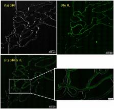
          Microfluidics is used increasingly for engineering and biomedical applications due to recent advances in microfabrication technologies. Visualization of bubbles, tracer particles, and cells in a microfluidic device is important for designing a device and analyzing results. However, with conventional methods, it is difficult to observe the channel geometry and such particles simultaneously. To overcome this limitation, we developed a Darkfield Internal Reflection Illumination (DIRI) system that improved the drawbacks of a conventional darkfield illuminator. This study was performed to investigate its utility in the field of microfluidics. The results showed that the developed system could clearly visualize both microbubbles and the channel wall by utilizing brightfield and DIRI illumination simultaneously. The methodology is useful not only for static phenomena, such as clogging, but also for dynamic phenomena, such as the detection of bubbles flowing in a channel. The system was also applied to simultaneous fluorescence and DIRI imaging. Fluorescent tracer beads and channel walls were observed clearly, which may be an advantage for future microparticle image velocimetry (μPIV) analysis, especially near a wall. Two types of cell stained with different colors, and the channel wall, can be recognized using the combined confocal and DIRI system. Whole-slide imaging was also conducted successfully using this system. The tiling function significantly expands the observing area of microfluidics. The developed system will be useful for a wide variety of engineering and biomedical applications for the growing field of microfluidics.

          Related collections

          Most cited references34

          • Record: found
          • Abstract: found
          • Article: not found

          Droplet microfluidics.

          Droplet-based microfluidic systems have been shown to be compatible with many chemical and biological reagents and capable of performing a variety of "digital fluidic" operations that can be rendered programmable and reconfigurable. This platform has dimensional scaling benefits that have enabled controlled and rapid mixing of fluids in the droplet reactors, resulting in decreased reaction times. This, coupled with the precise generation and repeatability of droplet operations, has made the droplet-based microfluidic system a potent high throughput platform for biomedical research and applications. In addition to being used as microreactors ranging from the nano- to femtoliter range; droplet-based systems have also been used to directly synthesize particles and encapsulate many biological entities for biomedicine and biotechnology applications. This review will focus on the various droplet operations, as well as the numerous applications of the system. Due to advantages unique to droplet-based systems, this technology has the potential to provide novel solutions to today's biomedical engineering challenges for advanced diagnostics and therapeutics.
            Bookmark
            • Record: found
            • Abstract: found
            • Article: not found

            Detection of mutations in EGFR in circulating lung-cancer cells.

            The use of tyrosine kinase inhibitors to target the epidermal growth factor receptor gene (EGFR) in patients with non-small-cell lung cancer is effective but limited by the emergence of drug-resistance mutations. Molecular characterization of circulating tumor cells may provide a strategy for noninvasive serial monitoring of tumor genotypes during treatment. We captured highly purified circulating tumor cells from the blood of patients with non-small-cell lung cancer using a microfluidic device containing microposts coated with antibodies against epithelial cells. We performed EGFR mutational analysis on DNA recovered from circulating tumor cells using allele-specific polymerase-chain-reaction amplification and compared the results with those from concurrently isolated free plasma DNA and from the original tumor-biopsy specimens. We isolated circulating tumor cells from 27 patients with metastatic non-small-cell lung cancer (median number, 74 cells per milliliter). We identified the expected EGFR activating mutation in circulating tumor cells from 11 of 12 patients (92%) and in matched free plasma DNA from 4 of 12 patients (33%) (P=0.009). We detected the T790M mutation, which confers drug resistance, in circulating tumor cells collected from patients with EGFR mutations who had received tyrosine kinase inhibitors. When T790M was detectable in pretreatment tumor-biopsy specimens, the presence of the mutation correlated with reduced progression-free survival (7.7 months vs. 16.5 months, P<0.001). Serial analysis of circulating tumor cells showed that a reduction in the number of captured cells was associated with a radiographic tumor response; an increase in the number of cells was associated with tumor progression, with the emergence of additional EGFR mutations in some cases. Molecular analysis of circulating tumor cells from the blood of patients with lung cancer offers the possibility of monitoring changes in epithelial tumor genotypes during the course of treatment. 2008 Massachusetts Medical Society
              Bookmark
              • Record: found
              • Abstract: found
              • Article: not found

              Formation of droplets and bubbles in a microfluidic T-junction-scaling and mechanism of break-up.

              This article describes the process of formation of droplets and bubbles in microfluidic T-junction geometries. At low capillary numbers break-up is not dominated by shear stresses: experimental results support the assertion that the dominant contribution to the dynamics of break-up arises from the pressure drop across the emerging droplet or bubble. This pressure drop results from the high resistance to flow of the continuous (carrier) fluid in the thin films that separate the droplet from the walls of the microchannel when the droplet fills almost the entire cross-section of the channel. A simple scaling relation, based on this assertion, predicts the size of droplets and bubbles produced in the T-junctions over a range of rates of flow of the two immiscible phases, the viscosity of the continuous phase, the interfacial tension, and the geometrical dimensions of the device.
                Bookmark

                Author and article information

                Contributors
                Role: Academic Editor
                Journal
                PLoS One
                PLoS ONE
                plos
                plosone
                PLoS ONE
                Public Library of Science (San Francisco, CA USA )
                1932-6203
                6 March 2015
                2015
                : 10
                : 3
                : e0116925
                Affiliations
                [1 ]The Department of Biomedical Engineering, Graduate School of Biomedical Engineering, Tohoku University, Sendai, Miyagi, Japan
                [2 ]Olympus Corporation, Shinjuku-Ku, Tokyo, Japan
                [3 ]Olympus Scientific Solutions Americas, Waltham, Massachusetts, United States of America
                [4 ]Olympus Engineering, Hachioji, Tokyo, Japan
                [5 ]Olympus Soft Imaging Solutions GmbH, Münster, Germany
                [6 ]Department of Bioengineering and Robotics, Tohoku University, Sendai, Miyagi, Japan
                University of Illinois at Chicago, UNITED STATES
                Author notes

                Competing Interests: Olympus America Inc. and Olympus Corporation provided microscope equipment but was not a funder of, and did not influence the outcome of, the study. The VS120 WSI scanning system was used. Yoshihiro Kawano and Chino Otsuka are employed by Olympus Corporation, James Sanzo and Christopher Higgins are employed by Olympus America, Tatsuo Nirei by Olympus Engineering and Tobias Schilling by Olympus Soft Imaging Solutions GmbH. This particular microscope setup (VS120 with BX-DSU) is not available in the market. There are no further patents, products in development or marketed products to declare. This does not alter the authors’ adherence to all the PLOS ONE policies on sharing data and materials, as detailed online in the guide for authors.

                Conceived and designed the experiments: YK CO JS CH TN TS. Performed the experiments: YK CO JS CH. Analyzed the data: YK CO JS CH TS TI. Contributed reagents/materials/analysis tools: YK CO JS. Wrote the paper: YK CO JS CH TS TI.

                Article
                PONE-D-14-32434
                10.1371/journal.pone.0116925
                4352060
                25748425
                597b6148-a490-4a07-9eb5-5ed7c19d5904
                Copyright @ 2015

                This is an open access article distributed under the terms of the Creative Commons Attribution License, which permits unrestricted use, distribution, and reproduction in any medium, provided the original author and source are credited

                History
                : 20 July 2014
                : 16 December 2014
                Page count
                Figures: 11, Tables: 0, Pages: 19
                Funding
                The authors used the following grant for their study: Grants-in-Aid for Scientific Research (A), No. 26242039, http://www.jsps.go.jp/english/e-grants/index.html. The funders had no role in study design, data collection and analysis, decision to publish, or preparation of the manuscript.
                Categories
                Research Article
                Custom metadata
                All relevant data are within the paper and its Supporting Information files.

                Uncategorized
                Uncategorized

                Comments

                Comment on this article