29
views
0
recommends
+1 Recommend
0 collections
    0
    shares
      • Record: found
      • Abstract: found
      • Article: found
      Is Open Access

      Ubiquitin-proteasome system controls ciliogenesis at the initial step of axoneme extension

      research-article

      Read this article at

      Bookmark
          There is no author summary for this article yet. Authors can add summaries to their articles on ScienceOpen to make them more accessible to a non-specialist audience.

          Abstract

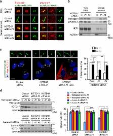
          Primary cilia are microtubule-based sensory organelles that organize numerous key signals during developments and tissue homeostasis. Ciliary microtubule doublet, named axoneme, is grown directly from the distal end of mother centrioles through a multistep process upon cell cycle exit; however, the instructive signals that initiate these events are poorly understood. Here we show that ubiquitin-proteasome machinery removes trichoplein, a negative regulator of ciliogenesis, from mother centrioles and thereby causes Aurora-A inactivation, leading to ciliogenesis. Ciliogenesis is blocked if centriolar trichoplein is stabilized by treatment with proteasome inhibitors or by expression of non-ubiquitylatable trichoplein mutant (K50/57R). Started from two-stepped global E3 screening, we have identified KCTD17 as a substrate-adaptor for Cul3-RING E3 ligases (CRL3s) that polyubiquitylates trichoplein. Depletion of KCTD17 specifically arrests ciliogenesis at the initial step of axoneme extension through aberrant trichoplein-Aurora-A activity. Thus, CRL3-KCTD17 targets trichoplein to proteolysis to initiate the axoneme extension during ciliogenesis.

          Abstract

          Biogenesis of the primary cilium begins after cell cycle exit, but the regulatory steps for its formation are poorly defined. Here the authors show that proteasome-mediated removal of the ciliogenesis inhibitor, trichoplein, from mother centrioles initiates the first step of ciliogenesis.

          Related collections

          Most cited references38

          • Record: found
          • Abstract: found
          • Article: not found

          Centrioles, centrosomes, and cilia in health and disease.

          Centrioles are barrel-shaped structures that are essential for the formation of centrosomes, cilia, and flagella. Here we review recent advances in our understanding of the function and biogenesis of these organelles, and we emphasize their connection to human disease. Deregulation of centrosome numbers has long been proposed to contribute to genome instability and tumor formation, whereas mutations in centrosomal proteins have recently been genetically linked to microcephaly and dwarfism. Finally, structural or functional centriole aberrations contribute to ciliopathies, a variety of complex diseases that stem from the absence or dysfunction of cilia.
            Bookmark
            • Record: found
            • Abstract: found
            • Article: not found

            Genome-Wide and Functional Annotation of Human E3 Ubiquitin Ligases Identifies MULAN, a Mitochondrial E3 that Regulates the Organelle's Dynamics and Signaling

            Specificity of protein ubiquitylation is conferred by E3 ubiquitin (Ub) ligases. We have annotated ∼617 putative E3s and substrate-recognition subunits of E3 complexes encoded in the human genome. The limited knowledge of the function of members of the large E3 superfamily prompted us to generate genome-wide E3 cDNA and RNAi expression libraries designed for functional screening. An imaging-based screen using these libraries to identify E3s that regulate mitochondrial dynamics uncovered MULAN/FLJ12875, a RING finger protein whose ectopic expression and knockdown both interfered with mitochondrial trafficking and morphology. We found that MULAN is a mitochondrial protein – two transmembrane domains mediate its localization to the organelle's outer membrane. MULAN is oriented such that its E3-active, C-terminal RING finger is exposed to the cytosol, where it has access to other components of the Ub system. Both an intact RING finger and the correct subcellular localization were required for regulation of mitochondrial dynamics, suggesting that MULAN's downstream effectors are proteins that are either integral to, or associated with, mitochondria and that become modified with Ub. Interestingly, MULAN had previously been identified as an activator of NF-κB, thus providing a link between mitochondrial dynamics and mitochondria-to-nucleus signaling. These findings suggest the existence of a new, Ub-mediated mechanism responsible for integration of mitochondria into the cellular environment.
              Bookmark
              • Record: found
              • Abstract: found
              • Article: not found

              Plk4-induced centriole biogenesis in human cells.

              We show that overexpression of Polo-like kinase 4 (Plk4) in human cells induces centrosome amplification through the simultaneous generation of multiple procentrioles adjoining each parental centriole. This provided an opportunity for dissecting centriole assembly and characterizing assembly intermediates. Critical components were identified and ordered into an assembly pathway through siRNA and localized through immunoelectron microscopy. Plk4, hSas-6, CPAP, Cep135, gamma-tubulin, and CP110 were required at different stages of procentriole formation and in association with different centriolar structures. Remarkably, hSas-6 associated only transiently with nascent procentrioles, whereas Cep135 and CPAP formed a core structure within the proximal lumen of both parental and nascent centrioles. Finally, CP110 was recruited early and then associated with the growing distal tips, indicating that centrioles elongate through insertion of alpha-/beta-tubulin underneath a CP110 cap. Collectively, these data afford a comprehensive view of the assembly pathway underlying centriole biogenesis in human cells.
                Bookmark

                Author and article information

                Journal
                Nat Commun
                Nat Commun
                Nature Communications
                Nature Pub. Group
                2041-1723
                01 October 2014
                : 5
                : 5081
                Affiliations
                [1 ]Division of Biochemistry, Aichi Cancer Center Research Institute , Nagoya, Aichi 464-8681, Japan
                [2 ]Department of Oncology, Graduate School of Pharmaceutical Sciences, Nagoya City University , Nagoya, Aichi 467-8603, Japan
                [3 ]Molecular Profiling Research Center for Drug Discovery, National Institute of Advanced Industrial Science and Technology , Tokyo 135-0064, Japan
                [4 ]Virology Division, National Cancer Center Research Institute , Tokyo 104-0045, Japan
                [5 ]Electron Microscope Laboratory, RIKEN Center for Developmental Biology , Kobe 650-0047, Japan
                [6 ]Japan Biological Informatics Consortium (JBiC) , Tokyo 135-8073, Japan
                [7 ]Laboratory of Cell Asymmetry, RIKEN Center of Developmental Biology , Kobe 650-0047, Japan
                [8 ]Department of Cellular Oncology, Nagoya University Graduate School of Medicine , Nagoya, Aichi 466-8550, Japan
                Author notes
                Article
                ncomms6081
                10.1038/ncomms6081
                4205846
                25270598
                5d38cc05-05c0-4a44-a4fe-3ea389f1c084
                Copyright © 2014, Nature Publishing Group, a division of Macmillan Publishers Limited. All Rights Reserved.

                This work is licensed under a Creative Commons Attribution 4.0 International License. The images or other third party material in this article are included in the article’s Creative Commons license, unless indicated otherwise in the credit line; if the material is not included under the Creative Commons license, users will need to obtain permission from the license holder to reproduce the material. To view a copy of this license, visit http://creativecommons.org/licenses/by/4.0/

                History
                : 22 April 2014
                : 27 August 2014
                Categories
                Article

                Uncategorized
                Uncategorized

                Comments

                Comment on this article