32
views
0
recommends
+1 Recommend
0 collections
    0
    shares
      • Record: found
      • Abstract: found
      • Article: not found

      Magnetic resonance imaging of brain angiogenesis after stroke

      research-article

      Read this article at

      ScienceOpenPublisherPMC
      Bookmark
          There is no author summary for this article yet. Authors can add summaries to their articles on ScienceOpen to make them more accessible to a non-specialist audience.

          Abstract

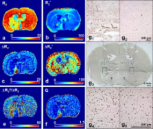
          Stroke is a major cause of mortality and long-term disability worldwide. The initial changes in local perfusion and tissue status underlying loss of brain function are increasingly investigated with noninvasive imaging methods. In addition, there is a growing interest in imaging of processes that contribute to post-stroke recovery. In this review, we discuss the application of magnetic resonance imaging (MRI) to assess the formation of new vessels by angiogenesis, which is hypothesized to participate in brain plasticity and functional recovery after stroke. The excellent soft tissue contrast, high spatial and temporal resolution, and versatility render MRI particularly suitable to monitor the dynamic processes involved in vascular remodeling after stroke. Here we review recent advances in the field of MR imaging that are aimed at assessment of tissue perfusion and microvascular characteristics, including cerebral blood flow and volume, vascular density, size and integrity. The potential of MRI to noninvasively monitor the evolution of post-ischemic angiogenic processes is demonstrated from a variety of in vivo studies in experimental stroke models. Finally, we discuss some pitfalls and limitations that may critically affect the accuracy and interpretation of MRI-based measures of (neo)vascularization after stroke.

          Related collections

          Most cited references65

          • Record: found
          • Abstract: found
          • Article: not found

          Susceptibility weighted imaging (SWI).

          Susceptibility differences between tissues can be utilized as a new type of contrast in MRI that is different from spin density, T1-, or T2-weighted imaging. Signals from substances with different magnetic susceptibilities compared to their neighboring tissue will become out of phase with these tissues at sufficiently long echo times (TEs). Thus, phase imaging offers a means of enhancing contrast in MRI. Specifically, the phase images themselves can provide excellent contrast between gray matter (GM) and white matter (WM), iron-laden tissues, venous blood vessels, and other tissues with susceptibilities that are different from the background tissue. Also, for the first time, projection phase images are shown to demonstrate tissue (vessel) continuity. In this work, the best approach for combining magnitude and phase images is discussed. The phase images are high-pass-filtered and then transformed to a special phase mask that varies in amplitude between zero and unity. This mask is multiplied a few times into the original magnitude image to create enhanced contrast between tissues with different susceptibilities. For this reason, this method is referred to as susceptibility-weighted imaging (SWI). Mathematical arguments are presented to determine the number of phase mask multiplications that should take place. Examples are given for enhancing GM/WM contrast and water/fat contrast, identifying brain iron, and visualizing veins in the brain. Copyright 2004 Wiley-Liss, Inc.
            Bookmark
            • Record: found
            • Abstract: found
            • Article: not found

            Repairing the human brain after stroke: I. Mechanisms of spontaneous recovery.

            Stroke remains a leading cause of adult disability. Some degree of spontaneous behavioral recovery is usually seen in the weeks after stroke onset. Variability in recovery is substantial across human patients. Some principles have emerged; for example, recovery occurs slowest in those destined to have less successful outcomes. Animal studies have extended these observations, providing insight into a broad range of underlying molecular and physiological events. Brain mapping studies in human patients have provided observations at the systems level that often parallel findings in animals. In general, the best outcomes are associated with the greatest return toward the normal state of brain functional organization. Reorganization of surviving central nervous system elements supports behavioral recovery, for example, through changes in interhemispheric lateralization, activity of association cortices linked to injured zones, and organization of cortical representational maps. A number of factors influence events supporting stroke recovery, such as demographics, behavioral experience, and perhaps genetics. Such measures gain importance when viewed as covariates in therapeutic trials of restorative agents that target stroke recovery.
              Bookmark
              • Record: found
              • Abstract: found
              • Article: not found

              High resolution measurement of cerebral blood flow using intravascular tracer bolus passages. Part I: Mathematical approach and statistical analysis.

              The authors review the theoretical basis of determination of cerebral blood flow (CBF) using dynamic measurements of nondiffusible contrast agents, and demonstrate how parametric and nonparametric deconvolution techniques can be modified for the special requirements of CBF determination using dynamic MRI. Using Monte Carlo modeling, the use of simple, analytical residue models is shown to introduce large errors in flow estimates when actual, underlying vascular characteristics are not sufficiently described by the chosen function. The determination of the shape of the residue function on a regional basis is shown to be possible only at high signal-to-noise ratio. Comparison of several nonparametric deconvolution techniques showed that a nonparametric deconvolution technique (singular value decomposition) allows estimation of flow relatively independent of underlying vascular structure and volume even at low signal-to-noise ratio associated with pixel-by-pixel deconvolution.
                Bookmark

                Author and article information

                Contributors
                +31-30-2535569 , +31-30-2535561 , P.Seevinck@umcutrecht.nl
                +31-30-2535569 , + 31-30-2535561 , rick@invivonmr.uu.nl
                Journal
                Angiogenesis
                Angiogenesis
                Springer Netherlands (Dordrecht )
                0969-6970
                1573-7209
                16 June 2010
                16 June 2010
                June 2010
                : 13
                : 2
                : 101-111
                Affiliations
                Biomedical MR Imaging and Spectroscopy Group, Image Sciences Institute, University Medical Center Utrecht, Yalelaan 2, 3584 CM Utrecht, The Netherlands
                Article
                9174
                10.1007/s10456-010-9174-0
                2911530
                20552268
                5d646315-03e5-4670-9532-241dcab9eff9
                © The Author(s) 2010
                History
                : 27 April 2010
                : 1 June 2010
                Categories
                Original Paper
                Custom metadata
                © Springer Science+Business Media B.V. 2010

                Human biology
                mri,microvessel size,angiogenesis,stroke,cerebral blood volume,brain ischemia,magnetic resonance imaging,cerebral blood flow,blood–brain barrier leakage,microvessel density

                Comments

                Comment on this article