59
views
0
recommends
+1 Recommend
0 collections
    0
    shares
      • Record: found
      • Abstract: found
      • Article: not found

      O-GlcNAc transferase invokes nucleotide sugar pyrophosphate participation in catalysis

      research-article

      Read this article at

      ScienceOpenPublisherPMC
      Bookmark
          There is no author summary for this article yet. Authors can add summaries to their articles on ScienceOpen to make them more accessible to a non-specialist audience.

          Abstract

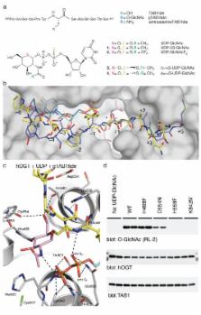
          Protein O-GlcNAcylation is an essential post-translational modification on hundreds of intracellular proteins in metazoa, catalyzed by O-GlcNAc transferase using unknown mechanisms of transfer and substrate recognition. Through crystallographic snapshots and mechanism-inspired chemical probes, we define how human O-GlcNAc transferase recognizes the sugar donor and acceptor peptide and employs a novel catalytic mechanism of glycosyl transfer, involving the sugar donor α-phosphate as the catalytic base, as well as an essential lysine. This mechanism appears to be a unique evolutionary solution to the spatial constraints imposed by a bulky protein acceptor substrate, and explains the unexpected specificity of a recently reported metabolic O-GlcNAc transferase inhibitor.

          Related collections

          Most cited references22

          • Record: found
          • Abstract: found
          • Article: not found

          Cross talk between O-GlcNAcylation and phosphorylation: roles in signaling, transcription, and chronic disease.

          O-GlcNAcylation is the addition of β-D-N-acetylglucosamine to serine or threonine residues of nuclear and cytoplasmic proteins. O-linked N-acetylglucosamine (O-GlcNAc) was not discovered until the early 1980s and still remains difficult to detect and quantify. Nonetheless, O-GlcNAc is highly abundant and cycles on proteins with a timescale similar to protein phosphorylation. O-GlcNAc occurs in organisms ranging from some bacteria to protozoans and metazoans, including plants and nematodes up the evolutionary tree to man. O-GlcNAcylation is mostly on nuclear proteins, but it occurs in all intracellular compartments, including mitochondria. Recent glycomic analyses have shown that O-GlcNAcylation has surprisingly extensive cross talk with phosphorylation, where it serves as a nutrient/stress sensor to modulate signaling, transcription, and cytoskeletal functions. Abnormal amounts of O-GlcNAcylation underlie the etiology of insulin resistance and glucose toxicity in diabetes, and this type of modification plays a direct role in neurodegenerative disease. Many oncogenic proteins and tumor suppressor proteins are also regulated by O-GlcNAcylation. Current data justify extensive efforts toward a better understanding of this invisible, yet abundant, modification. As tools for the study of O-GlcNAc become more facile and available, exponential growth in this area of research will eventually take place.
            Bookmark
            • Record: found
            • Abstract: found
            • Article: not found

            Structure of human O-GlcNAc transferase and its complex with a peptide substrate

            O-GlcNAc transferase (OGT) is an essential mammalian enzyme that couples metabolic status to the regulation of a wide variety of cellular signaling pathways by acting as a nutrient sensor1. OGT catalyzes the transfer of N-acetyl-glucosamine from UDP-GlcNAc to serines and threonines of cytoplasmic, nuclear and mitochondrial proteins2,3, including numerous transcription factors4, tumor suppressors, kinases5, phosphatases1, and histone-modifying proteins6. Aberrant O-GlcNAcylation by OGT has been linked to insulin resistance7, diabetic complications8, cancer9 and neurodegenerative diseases including Alzheimer’s10. Despite the importance of OGT, the details of how it recognizes and glycosylates its protein substrates are largely unknown. We report here two crystal structures of human OGT, as a binary complex with UDP (2.8 A) and a ternary complex with UDP and a peptide substrate (1.95 A). The structures provide clues to the enzyme mechanism, show how OGT recognizes target peptide sequences, and reveal the fold of the unique domain between the two halves of the catalytic region. This information will accelerate the rational design of biological experiments to investigate OGT’s functions and the design of inhibitors for use as cellular probes and to assess its potential as a therapeutic target.
              Bookmark
              • Record: found
              • Abstract: found
              • Article: not found

              Structural basis of perturbed pKa values of catalytic groups in enzyme active sites.

              In protein and RNA macromolecules, only a limited number of different side-chain chemical groups are available to function as catalysts. The myriad of enzyme-catalyzed reactions results from the ability of most of these groups to function either as nucleophilic, electrophilic, or general acid-base catalysts, and the key to their adapted chemical function lies in their states of protonation. Ionization is determined by the intrinsic pKa of the group and the microenvironment created around the group by the protein or RNA structure, which perturbs its intrinsic pKa to its functional or apparent pKa. These pKa shifts result from interactions of the catalytic group with other fully or partially charged groups as well as the polarity or dielectric of the medium that surrounds it. The electrostatic interactions between ionizable groups found on the surface of macromolecules are weak and cause only slight pKa perturbations ( 2 units) and are the subject of this review. The magnitudes of these pKa perturbations are analyzed with respect to the structural details of the active-site microenvironment and the energetics of the reactions that they catalyze.
                Bookmark

                Author and article information

                Journal
                101231976
                32624
                Nat Chem Biol
                Nat. Chem. Biol.
                Nature chemical biology
                1552-4450
                1552-4469
                9 October 2012
                28 October 2012
                December 2012
                01 June 2013
                : 8
                : 12
                : 969-974
                Affiliations
                [1 ]College of Life Sciences, University of Dundee, Dundee, UK
                [2 ]Louisiana State University, Baton Rouge, LA, USA
                Author notes
                [†]

                These authors contributed equally to this work

                [* ]Correspondence to: dmfvanaalten@ 123456dundee.ac.uk

                Author Contributions M.S. and D.M.F.v.A., structural biology; M.S. and X.Z., protein expression and enzyme activity assays; V.S.B., synthetic organic chemistry; D.E.B., enzyme kinetics measurements; I.N., X.Z., D.A.R. and T.A., surface plasmon resonance experiments; A.F., molecular biology; O.A., mass spectrometry. M.S., X.Z., V.S.B. and D.M.F.v.A. devised the experiments, M.S., X.Z., V.S.B., M.A.M., A.W.S. and D.M.F.v.A. interpreted the data and wrote the manuscript.

                Article
                EMS50081
                10.1038/nchembio.1108
                3509171
                23103942
                5e9a3963-712c-419a-8e60-1cb9c9328503

                Users may view, print, copy, download and text and data- mine the content in such documents, for the purposes of academic research, subject always to the full Conditions of use: http://www.nature.com/authors/editorial_policies/license.html#terms

                History
                Funding
                Funded by: Wellcome Trust :
                Award ID: 087590 || WT
                Categories
                Article

                Biochemistry
                Biochemistry

                Comments

                Comment on this article