2
views
0
recommends
+1 Recommend
0 collections
    0
    shares
      • Record: found
      • Abstract: found
      • Article: found
      Is Open Access

      Fluorescent Nanodiamonds Enable Long-Term Detection of Human Adipose-Derived Stem/Stromal Cells in an In Vivo Chondrogenesis Model Using Decellularized Extracellular Matrices and Fibrin Glue Polymer

      research-article

      Read this article at

      Bookmark
          There is no author summary for this article yet. Authors can add summaries to their articles on ScienceOpen to make them more accessible to a non-specialist audience.

          Abstract

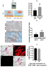
          Clinically available materials, including allogeneic irradiated costal cartilage and fibrin glue polymer, were used as scaffolds for in vivo chondrogenic differentiation of human adipose-derived stem/stromal cells (hASCs) in the attempt to develop a more efficient treatment over current methods. Current studies include the use of growth-factor stimulation, tissue engineering, and biocompatible materials; however, most methods involve complicated processes and pose clinical limitations. In this report, the xenografts in the experimental group composed of a diced decellularized cartilage extracellular matrix (ECM), hASCs, and fibrin glue polymer were implanted into the subcutaneous layer of nude mice, and the results were compared with two groups of controls; one control group received implantation of decellularized cartilage ECM and fibrin glue polymer, and the other control group received implantation of hASCs mixed with fibrin glue polymer. To evaluate whether hASCs had in vivo chondrogenesis in the xenografts, hASCs were labeled with fluorescent nanodiamonds (FNDs), a biocompatible and photostable nanomaterial, to allow for long-term detection and histological analysis. Increased cellularity, glycosaminoglycan, and collagen deposition were found by the histological examination in the experimental group compared with control groups. With the background-free detection technique and time-gated fluorescence imaging, the numbers and locations of the FND-labeled hASCs could be detected by confocal microscopy. The chondrocyte-specific markers, such as aggrecan and type II collagen, were colocalized with cells containing signals of FNDs which indicated in vivo chondrogenesis of hASCs. Taken together, functional in vivo chondrogenesis of the hASCs could be achieved by clinically available decellularized cartilage ECM and fibrin glue polymer in the nude mice model without in vitro chondrogenic induction. The fluorescent signals of FNDs in hASCs can be detected in histological analysis, such as hematoxylin and eosin staining (H&E staining) without the interference of the autofluorescence. Our study may warrant future clinical applications of the combination of decellular cartilage ECM, fibrin glue polymer, and hASCs for cartilage repair.

          Related collections

          Most cited references33

          • Record: found
          • Abstract: found
          • Article: not found

          The properties and applications of nanodiamonds.

          Nanodiamonds have excellent mechanical and optical properties, high surface areas and tunable surface structures. They are also non-toxic, which makes them well suited to biomedical applications. Here we review the synthesis, structure, properties, surface chemistry and phase transformations of individual nanodiamonds and clusters of nanodiamonds. In particular we discuss the rational control of the mechanical, chemical, electronic and optical properties of nanodiamonds through surface doping, interior doping and the introduction of functional groups. These little gems have a wide range of potential applications in tribology, drug delivery, bioimaging and tissue engineering, and also as protein mimics and a filler material for nanocomposites.
            Bookmark
            • Record: found
            • Abstract: found
            • Article: not found

            A controlled trial of arthroscopic surgery for osteoarthritis of the knee.

            Many patients report symptomatic relief after undergoing arthroscopy of the knee for osteoarthritis, but it is unclear how the procedure achieves this result. We conducted a randomized, placebo-controlled trial to evaluate the efficacy of arthroscopy for osteoarthritis of the knee. A total of 180 patients with osteoarthritis of the knee were randomly assigned to receive arthroscopic débridement, arthroscopic lavage, or placebo surgery. Patients in the placebo group received skin incisions and underwent a simulated débridement without insertion of the arthroscope. Patients and assessors of outcome were blinded to the treatment-group assignment. Outcomes were assessed at multiple points over a 24-month period with the use of five self-reported scores--three on scales for pain and two on scales for function--and one objective test of walking and stair climbing. A total of 165 patients completed the trial. At no point did either of the intervention groups report less pain or better function than the placebo group. For example, mean (+/-SD) scores on the Knee-Specific Pain Scale (range, 0 to 100, with higher scores indicating more severe pain) were similar in the placebo, lavage, and débridement groups: 48.9+/-21.9, 54.8+/-19.8, and 51.7+/-22.4, respectively, at one year (P=0.14 for the comparison between placebo and lavage; P=0.51 for the comparison between placebo and débridement) and 51.6+/-23.7, 53.7+/-23.7, and 51.4+/-23.2, respectively, at two years (P=0.64 and P=0.96, respectively). Furthermore, the 95 percent confidence intervals for the differences between the placebo group and the intervention groups exclude any clinically meaningful difference. In this controlled trial involving patients with osteoarthritis of the knee, the outcomes after arthroscopic lavage or arthroscopic débridement were no better than those after a placebo procedure.
              Bookmark
              • Record: found
              • Abstract: found
              • Article: not found

              Mass production and dynamic imaging of fluorescent nanodiamonds.

              Fluorescent nanodiamond is a new nanomaterial that possesses several useful properties, including good biocompatibility, excellent photostability and facile surface functionalizability. Moreover, when excited by a laser, defect centres within the nanodiamond emit photons that are capable of penetrating tissue, making them well suited for biological imaging applications. Here, we show that bright fluorescent nanodiamonds can be produced in large quantities by irradiating synthetic diamond nanocrystallites with helium ions. The fluorescence is sufficiently bright and stable to allow three-dimensional tracking of a single particle within the cell by means of either one- or two-photon-excited fluorescence microscopy. The excellent photophysical characteristics are maintained for particles as small as 25 nm, suggesting that fluorescent nanodiamond is an ideal probe for long-term tracking and imaging in vivo, with good temporal and spatial resolution.
                Bookmark

                Author and article information

                Journal
                Polymers (Basel)
                Polymers (Basel)
                polymers
                Polymers
                MDPI
                2073-4360
                23 August 2019
                September 2019
                : 11
                : 9
                : 1391
                Affiliations
                [1 ]Institute of Stem Cell and Translational Cancer Research, Chang Gung Memorial Hospital at Linko, Taoyuan 333, Taiwan
                [2 ]Ph.D. Program in Translational Medicine, Kaohsiung Medical University, Kaohsiung, and Academia Sinica, Taipei 115, Taiwan
                [3 ]Regeneration Medicine and Cell Therapy Research Center, Kaohsiung Medical University, Kaohsiung 807 Taiwan
                [4 ]Division of Plastic Surgery, Department of Surgery, Kaohsiung Medical University Hospital, Kaohsiung 807, Taiwan
                [5 ]Center of Teaching and Research, Kaohsiung Municipal Ta-Tung Hospital, Kaohsiung 801, Taiwan
                [6 ]Graduate Institute of Biomedical Engineering, National Chung Hsing University, Taichung 402, Taiwan
                [7 ]Institute of Atomic and Molecular Sciences, Academia Sinica, Taipei 106, Taiwan
                [8 ]Orthopaedic Research Center, Kaohsiung Medical University, Kaohsiung 801, Taiwan
                [9 ]Institute of Cellular and Organismic Biology, Academia Sinica, Taipei 115, Taiwan
                Author notes
                [* ]Correspondence: johnyu@ 123456gate.sinica.edu.tw ; Tel.: +886-3-328-1200#7803
                Author information
                https://orcid.org/0000-0002-5162-9752
                https://orcid.org/0000-0001-8327-6835
                Article
                polymers-11-01391
                10.3390/polym11091391
                6780225
                31450801
                5eda9c7b-7db4-4b20-9d0c-55e8d72e7770
                © 2019 by the authors.

                Licensee MDPI, Basel, Switzerland. This article is an open access article distributed under the terms and conditions of the Creative Commons Attribution (CC BY) license ( http://creativecommons.org/licenses/by/4.0/).

                History
                : 16 June 2019
                : 16 August 2019
                Categories
                Article

                human adipose-derived stem/stromal cells (hascs),in vivo chondrogenesis,decellularized cartilage ecm,fibrin glue polymer,fluorescent nanodiamonds (fnds),long-term detection

                Comments

                Comment on this article