6
views
0
recommends
+1 Recommend
0 collections
    0
    shares
      • Record: found
      • Abstract: found
      • Article: found
      Is Open Access

      Time since menopause and skeletal muscle estrogen receptors, PGC-1α, and AMPK

      research-article

      Read this article at

      Bookmark
          There is no author summary for this article yet. Authors can add summaries to their articles on ScienceOpen to make them more accessible to a non-specialist audience.

          Abstract

          Objective:

          Short-term administration of estradiol (E 2) improves insulin-stimulated glucose disposal rate in early postmenopausal (EPM) women compared with a reduction in late postmenopausal (LPM) women. The underlying mechanisms by which E 2 action on glucose disposal rate reversed from beneficial early to harmful late in menopause is unknown, but might include adverse changes in estrogen receptors (ERs) or other biomarkers of cellular energy metabolism with age or duration of estrogen deficiency.

          Methods:

          We retrospectively analyzed skeletal muscle samples from 27 postmenopausal women who were 6 years or less past menopause (EPM; n = 13) or at least 10 years past menopause (LPM; n = 14). Fasted skeletal muscle (vastus lateralis) samples were collected after 1 week administration of transdermal E 2 or placebo, in random cross-over design.

          Results:

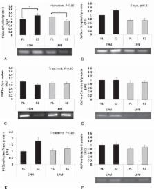
          Compared with EPM, LPM had reduced skeletal muscle ERα and ERβ nuclear protein. Short-term E 2 treatment did not change nuclear ERα or ERβ, but decreased cytosolic ERα, so the proportion of ERα in the nucleus compared with the cytosol tended to increase. There was a group-by-treatment interaction ( P < 0.05) for nuclear proliferator-activated receptor γ co-activator 1-α and phosphorylated adenosine monophosphate-activated protein kinase, such that E 2 increased these proteins in EPM, but decreased these proteins in LPM.

          Conclusions:

          These preliminary studies of skeletal muscle from early and late postmenopausal women treated with E 2 suggest there may be declines in skeletal muscle ER and changes in the E 2-mediated regulation of cellular energy homeostasis with increasing time since menopause.

          Related collections

          Most cited references28

          • Record: found
          • Abstract: found
          • Article: not found

          PGC-1alpha: a key regulator of energy metabolism.

          Peroxisome proliferator-activated receptor-gamma coactivator (PGC)-1alpha is a member of a family of transcription coactivators that plays a central role in the regulation of cellular energy metabolism. It is strongly induced by cold exposure, linking this environmental stimulus to adaptive thermogenesis. PGC-1alpha stimulates mitochondrial biogenesis and promotes the remodeling of muscle tissue to a fiber-type composition that is metabolically more oxidative and less glycolytic in nature, and it participates in the regulation of both carbohydrate and lipid metabolism. It is highly likely that PGC-1alpha is intimately involved in disorders such as obesity, diabetes, and cardiomyopathy. In particular, its regulatory function in lipid metabolism makes it an inviting target for pharmacological intervention in the treatment of obesity and Type 2 diabetes.
            Bookmark
            • Record: found
            • Abstract: found
            • Article: not found

            Regulation of mitochondrial biogenesis.

            Although it is well established that physical activity increases mitochondrial content in muscle, the molecular mechanisms underlying this process have only recently been elucidated. Mitochondrial dysfunction is an important component of different diseases associated with aging, such as Type 2 diabetes and Alzheimer's disease. PGC-1alpha (peroxisome-proliferator-activated receptor gamma co-activator-1alpha) is a co-transcriptional regulation factor that induces mitochondrial biogenesis by activating different transcription factors, including nuclear respiratory factor 1 and nuclear respiratory factor 2, which activate mitochondrial transcription factor A. The latter drives transcription and replication of mitochondrial DNA. PGC-1alpha itself is regulated by several different key factors involved in mitochondrial biogenesis, which will be reviewed in this chapter. Of those, AMPK (AMP-activated protein kinase) is of major importance. AMPK acts as an energy sensor of the cell and works as a key regulator of mitochondrial biogenesis. AMPK activity has been shown to decrease with age, which may contribute to decreased mitochondrial biogenesis and function with aging. Given the potentially important role of mitochondrial dysfunction in the pathogenesis of numerous diseases and in the process of aging, understanding the molecular mechanisms regulating mitochondrial biogenesis and function may provide potentially important novel therapeutic targets.
              Bookmark
              • Record: found
              • Abstract: found
              • Article: not found

              The emergence of the metabolic syndrome with menopause.

              Women with the metabolic syndrome (central obesity, insulin resistance, and dyslipidemia) are known to be at especially high risk for cardiovascular disease (CVD). The prevalence of the metabolic syndrome increases with menopause and may partially explain the apparent acceleration in CVD after menopause. The transition from pre- to postmenopause is associated with the emergence of many features of the metabolic syndrome, including 1) increased central (intraabdominal) body fat; 2) a shift toward a more atherogenic lipid profile, with increased low density lipoprotein and triglycerides levels, reduced high density lipoprotein, and small, dense low density lipoprotein particles; 3) and increased glucose and insulin levels. The emergence of these risk factors may be a direct result of ovarian failure or, alternatively, an indirect result of the metabolic consequences of central fat redistribution with estrogen deficiency. It is unclear whether the transition to menopause increases CVD risk in all women or only those who develop features of the metabolic syndrome. This article will review the features of the metabolic syndrome that emerge with estrogen deficiency. A better understanding of these metabolic changes with menopause will aid in the recognition and treatment of women at risk for future CVD, leading to appropriate interventions.
                Bookmark

                Author and article information

                Journal
                Menopause
                Menopause
                MENOP
                Menopause (New York, N.y.)
                Lippincott-Raven Publishers
                1072-3714
                1530-0374
                July 2017
                26 June 2017
                : 24
                : 7
                : 815-823
                Affiliations
                [1 ]Division of Geriatric Medicine
                [2 ]Division of Endocrinology, Diabetes and Metabolism, Department of Medicine, University of Colorado Anschutz Medical Campus, Aurora, CO
                [3 ]VA Eastern Colorado Health Care System, Geriatric Research Education and Clinical Center (GRECC), Denver, CO
                [4 ]Denver Health and Hospital Authority, Denver, CO
                [5 ]School of Kinesiology, University of Minnesota, Minneapolis, MN.
                Author notes
                Address correspondence to: Rachael E. Van Pelt, PhD, Division of Geriatric Medicine, University of Colorado Anschutz Medical Campus, 12631 East 17th Ave, Campus Box B-179, Aurora, CO 80045. E-mail: rachael.vanpelt@ 123456ucdenver.edu
                Article
                MENO-D-16-00260
                10.1097/GME.0000000000000829
                5484730
                28195989
                5f3e304d-142a-4f3e-a1d6-738200b35c22
                © 2017 The Author(s). Published by Wolters Kluwer Health, Inc. on behalf of The North American Menopause Society.

                This is an open access article distributed under the terms of the Creative Commons Attribution-Non Commercial-No Derivatives License 4.0 (CCBY-NC-ND), where it is permissible to download and share the work provided it is properly cited. The work cannot be changed in any way or used commercially without permission from the journal. http://creativecommons.org/licenses/by-nc-nd/4.0

                History
                : 08 August 2016
                : 06 December 2016
                : 06 December 2016
                Categories
                Original Articles
                Custom metadata
                TRUE

                ampk,estradiol,estrogen receptors,menopause,pgc-1α,skeletal muscle

                Comments

                Comment on this article