39
views
0
recommends
+1 Recommend
0 collections
    0
    shares
      • Record: found
      • Abstract: found
      • Article: found
      Is Open Access

      Stimulation of Midbrain Dopaminergic Structures Modifies Firing Rates of Rat Lateral Habenula Neurons

      research-article
      , , *
      PLoS ONE
      Public Library of Science

      Read this article at

      Bookmark
          There is no author summary for this article yet. Authors can add summaries to their articles on ScienceOpen to make them more accessible to a non-specialist audience.

          Abstract

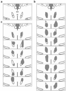
          Ventral tegmental area (VTA) and substantia nigra pars compacta (SNpc) are midbrain structures known to be involved in mediating reward in rodents. Lateral habenula (LHb) is considered as a negative reward source and it is reported that stimulation of the LHb rapidly induces inhibition of firing in midbrain dopamine neurons. Interestingly, the phasic fall in LHb neuronal activity may follow the excitation of dopamine neurons in response to reward-predicting stimuli. The VTA and SNpc give rise to dopaminergic projections that innervate the LHb, which is also known to be involved in processing painful stimuli. But it's unclear what physiological effects these inputs have on habenular function. In this study we distinguished the LHb pain-activated neurons of the Wistar rats and assessed their electrophysiological responsiveness to the stimulation of the VTA and SNpc with either single-pulse stimulation (300 µA, 0.5 Hz) or tetanic stimulation (80 µA, 25 Hz). Single-pulse stimulation that was delivered to either midbrain structure triggered transient inhibition of firing of ∼90% of the LHb pain-activated neurons. However, tetanic stimulation of the VTA tended to evoke an elevation in neuronal firing rate. We conclude that LHb pain-activated neurons can receive diverse reward-related signals originating from midbrain dopaminergic structures, and thus participate in the regulation of the brain reward system via both positive and negative feedback mechanisms.

          Related collections

          Most cited references31

          • Record: found
          • Abstract: found
          • Article: not found

          Dopamine reward circuitry: two projection systems from the ventral midbrain to the nucleus accumbens-olfactory tubercle complex.

          Anatomical and functional refinements of the meso-limbic dopamine system of the rat are discussed. Present experiments suggest that dopaminergic neurons localized in the posteromedial ventral tegmental area (VTA) and central linear nucleus raphe selectively project to the ventromedial striatum (medial olfactory tubercle and medial nucleus accumbens shell), whereas the anteromedial VTA has few if any projections to the ventral striatum, and the lateral VTA largely projects to the ventrolateral striatum (accumbens core, lateral shell and lateral tubercle). These findings complement the recent behavioral findings that cocaine and amphetamine are more rewarding when administered into the ventromedial striatum than into the ventrolateral striatum. Drugs such as nicotine and opiates are more rewarding when administered into the posterior VTA or the central linear nucleus than into the anterior VTA. A review of the literature suggests that (1) the midbrain has corresponding zones for the accumbens core and medial shell; (2) the striatal portion of the olfactory tubercle is a ventral extension of the nucleus accumbens shell; and (3) a model of two dopamine projection systems from the ventral midbrain to the ventral striatum is useful for understanding reward function. The medial projection system is important in the regulation of arousal characterized by affect and drive and plays a different role in goal-directed learning than the lateral projection system, as described in the variation-selection hypothesis of striatal functional organization.
            Bookmark
            • Record: found
            • Abstract: found
            • Article: not found

            D1 and D2 dopamine-receptor modulation of striatal glutamatergic signaling in striatal medium spiny neurons.

            Dopamine shapes a wide variety of psychomotor functions. This is mainly accomplished by modulating cortical and thalamic glutamatergic signals impinging upon principal medium spiny neurons (MSNs) of the striatum. Several lines of evidence suggest that dopamine D1 receptor signaling enhances dendritic excitability and glutamatergic signaling in striatonigral MSNs, whereas D2 receptor signaling exerts the opposite effect in striatopallidal MSNs. The functional antagonism between these two major striatal dopamine receptors extends to the regulation of synaptic plasticity. Recent studies, using transgenic mice in which cells express D1 and D2 receptors, have uncovered unappreciated differences between MSNs that shape glutamatergic signaling and the influence of DA on synaptic plasticity. These studies have also shown that long-term alterations in dopamine signaling produce profound and cell-type-specific reshaping of corticostriatal connectivity and function.
              Bookmark
              • Record: found
              • Abstract: found
              • Article: not found

              Opponent appetitive-aversive neural processes underlie predictive learning of pain relief.

              Termination of a painful or unpleasant event can be rewarding. However, whether the brain treats relief in a similar way as it treats natural reward is unclear, and the neural processes that underlie its representation as a motivational goal remain poorly understood. We used fMRI (functional magnetic resonance imaging) to investigate how humans learn to generate expectations of pain relief. Using a pavlovian conditioning procedure, we show that subjects experiencing prolonged experimentally induced pain can be conditioned to predict pain relief. This proceeds in a manner consistent with contemporary reward-learning theory (average reward/loss reinforcement learning), reflected by neural activity in the amygdala and midbrain. Furthermore, these reward-like learning signals are mirrored by opposite aversion-like signals in lateral orbitofrontal cortex and anterior cingulate cortex. This dual coding has parallels to 'opponent process' theories in psychology and promotes a formal account of prediction and expectation during pain.
                Bookmark

                Author and article information

                Contributors
                Role: Editor
                Journal
                PLoS One
                PLoS ONE
                plos
                plosone
                PLoS ONE
                Public Library of Science (San Francisco, USA )
                1932-6203
                2012
                2 April 2012
                : 7
                : 4
                : e34323
                Affiliations
                [1]Department of Physiology, Norman Bethune College of Medicine, Jilin University, Changchun, Jilin, China
                University of Chicago, United States of America
                Author notes

                Conceived and designed the experiments: HZ XFS. Performed the experiments: XFS XGR. Analyzed the data: XFS HZ. Contributed reagents/materials/analysis tools: HZ. Wrote the paper: XFS HZ.

                Article
                PONE-D-11-23480
                10.1371/journal.pone.0034323
                3317773
                22485164
                5fbd260a-7ee9-4fcb-b5a0-6410fd2ce9c3
                Shen et al. This is an open-access article distributed under the terms of the Creative Commons Attribution License, which permits unrestricted use, distribution, and reproduction in any medium, provided the original author and source are credited.
                History
                : 25 November 2011
                : 28 February 2012
                Page count
                Pages: 9
                Categories
                Research Article
                Biology
                Anatomy and Physiology
                Biochemistry
                Neurochemistry
                Computational Biology
                Computational Neuroscience
                Model Organisms
                Animal Models
                Neuroscience
                Cognitive Neuroscience
                Medicine

                Uncategorized
                Uncategorized

                Comments

                Comment on this article