12
views
0
recommends
+1 Recommend
0 collections
    0
    shares
      • Record: found
      • Abstract: found
      • Article: found
      Is Open Access

      Colon Preneoplastic Lesions in Animal Models

      review-article

      Read this article at

      Bookmark
          There is no author summary for this article yet. Authors can add summaries to their articles on ScienceOpen to make them more accessible to a non-specialist audience.

          Abstract

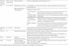
          The animal model is a powerful and fundamental tool in the field of biochemical research including toxicology, carcinogenesis, cancer therapeutics and prevention. In the carcinogenesis animal model system, numerous examples of preneoplastic lesions have been isolated and investigated from various perspectives. This may indicate that several options of endpoints to evaluate carcinogenesis effect or therapeutic outcome are presently available; however, classification of preneoplastic lesions has become complicated. For instance, these lesions include aberrant crypt foci (ACF), dysplastic ACF, flat ACF, β-catenin accumulated crypts, and mucin-depleted foci. These lesions have been induced by commonly used chemical carcinogens such as azoxymethane (AOM), 1,2-dimethylhydrazine (DMH), methylnitrosourea (MUN), or 2-amino-1-methyl-6-phenylimidazo[4,5- b]pyridine (PhIP). Investigators can choose any procedures or methods to examine colonic preneoplastic lesions according to their interests and the objectives of their experiments. Based on topographical, histopathological, and biological features of colon cancer preneoplastic lesions in the animal model, we summarize and discuss the character and implications of these lesions.

          Related collections

          Most cited references48

          • Record: found
          • Abstract: found
          • Article: not found

          Observation and quantification of aberrant crypts in the murine colon treated with a colon carcinogen: preliminary findings.

          R P Bird (1987)
          In the present study a methodological approach is taken which quantitates aberrant dysplastic crypts in the unsectioned murine colon. C57BL/6J or CF1 female mice (7-8 weeks old) were injected (i.p.) with azoxymethane (5 mg/kg body wt./week) for 4 weeks. Their colons were excised, cut open on the median axis and fixed flat in buffered formalin. Unsectioned colons were stained with methylene blue. The mucosal side was examined under a light microscope. The aberrant crypts, which are larger and have a thicker epithelial lining, were easily visualized using X 4 or X 10 objectives. CF1 mice, which are more sensitive to developing colon tumors, had a higher number of aberrant crypts/colon than their less sensitive counterparts, C57BL/6J mice (5.0 +/- 0.7 vs. 2.4 +/- 0.7). The usefulness of this observation as a possible measure of neoplastic events is discussed in the animal and human situation.
            Bookmark
            • Record: found
            • Abstract: found
            • Article: not found

            Role of aberrant crypt foci in understanding the pathogenesis of colon cancer.

            Aberrant crypt foci (ACF) are present in carcinogen treated rodent colons and in the colons of humans with a high risk for developing the disease. It is proposed that ACF are preneoplastic lesions. Quantification of the number and growth features of ACF has been employed to study modulators of colon carcinogenesis. In this review, examples are presented to support the concept that ACF are preneoplastic lesions and that sequential quantification of their number and growth features (crypt multiplicity) in animal colons may provide further insight into the pathogenesis of colon cancer. It is proposed that cellular and molecular heterogeneity among ACF with different growth and morphologic features will be invaluable in the identification of events critically associated with cancer development.
              Bookmark
              • Record: found
              • Abstract: found
              • Article: not found

              Peroxisome proliferator-activated receptor gamma ligands suppress colon carcinogenesis induced by azoxymethane in mice.

              Peroxisome proliferator-activated receptor gamma (PPARgamma) is known to regulate growth arrest and terminal differentiation of adipocytes and is used clinically as a new class of antidiabetic drugs. Recently, several studies have reported that treatment of cancer cells with PPARgamma ligands induces cell differentiation and apoptosis, suggesting a potential application as chemopreventive agents against carcinogenesis. However, contradictory results have been reported with regards to the biologic role of PPARgamma in carcinogenesis. Tanaka et al.(24) have recently reported the suppressive effect of a PPARgamma ligand, troglitazone (Tro), on the formation of aberrant crypt foci (ACF) in rats. In the present study, 3 different kinds of PPARgamma ligands were subjected to the experiments to confirm their suppressive effects on colon carcinogenesis. Three PPARgamma ligands, pioglitazone (Pio) (200 ppm), rosiglitazone (Rosi) (200 ppm), and Tro (1000 ppm) were investigated on the induction of ACF, a putative precancerous lesion of the colon, and colon tumor formation using an azoxymethane (AOM)-induced colon cancer model in BALB/c mice, and dose dependency of a PPARgamma ligand was also examined. PPARgamma ligands reduced the ACF formation by AOM (10 mg/kg body weight) and induction of colon tumors were also markedly suppressed by a continuous feeding of Pio at 200 ppm. Our findings indicate that PPARgamma ligands are indeed potential chemopreventive agents for colon carcinogenesis.
                Bookmark

                Author and article information

                Journal
                J Toxicol Pathol
                J Toxicol Pathol
                TOX
                Journal of Toxicologic Pathology
                Japanese Society of Toxicologic Pathology
                0914-9198
                1881-915X
                26 December 2013
                December 2013
                : 26
                : 4
                : 335-341
                Affiliations
                [1 ]Department of Molecular Toxicology, Graduate School of Medical Sciences and Medical School, Nagoya City University,1 Kawasumi, Mizuho-ku, Mizuho-cho, Nagoya 467-8601, Japan
                [2 ]Radiation Effect Accumulation and Prevention Project, Fukushima Project Headquarters and Radiobiology for Children’s Health Program, Research Center for Radiation Protection, National Institute of Radiological Sciences, 4-9-1 Anagawa, Inage-ku, Chiba 263-8555, Japan
                [3 ]Department of Pathology and Oncology, Graduate School of Medicine and Faculty of Medicine, University of the Ryukyus Faculty of Medicine, 207 Uehara, Nishihara-cho, Okinawa 903-0215, Japan
                Author notes
                *Corresponding author: M Suzui (e-mail: suzui@ 123456med.nagoya-cu.ac.jp )
                Article
                2013-0028
                10.1293/tox.2013-0028
                3921915
                24526805
                614b8846-dbf8-40a5-9dee-d17d3efc9ea5
                ©2013 The Japanese Society of Toxicologic Pathology

                This is an open-access article distributed under the terms of the Creative Commons Attribution Non-Commercial No Derivatives (by-nc-nd) License.

                History
                : 28 May 2013
                : 27 June 2013
                Categories
                Review

                Pathology
                preneoplastic lesion,colon carcinogenesis,animal model,topographic view
                Pathology
                preneoplastic lesion, colon carcinogenesis, animal model, topographic view

                Comments

                Comment on this article