15
views
0
recommends
+1 Recommend
0 collections
    0
    shares
      • Record: found
      • Abstract: found
      • Article: found
      Is Open Access

      Granulomatous/sarcoid-like lesions associated with checkpoint inhibitors: a marker of therapy response in a subset of melanoma patients

      case-report

      Read this article at

      Bookmark
          There is no author summary for this article yet. Authors can add summaries to their articles on ScienceOpen to make them more accessible to a non-specialist audience.

          Abstract

          Background

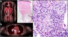
          Immune checkpoint therapy has dramatically changed the landscape of cancer therapy, providing an efficacious and durable therapeutic option for patients with advanced-stage disease. However, dermatologic toxicities are a well-recognized side effect in patients receiving this therapy. A spectrum of immune related adverse events (irAEs) involving the skin can occur and include immunobullous disorders, lichenoid dermatitis, and vitiligo. Granulomatous/sarcoid-like lesions are now being recognized with the current class of checkpoint inhibitors (CPIs) that involve the dermis, the subcutaneous tissue (panniculitis), and lymph nodes.

          Case presentation

          We report 3 patients who developed granulomatous/sarcoid-like lesions while being treated with immune checkpoint therapy for advanced-stage melanoma, and we provide a comprehensive review of the literature in which similar cases are described. To date, 26 patients (including the 3 from this report) have been described with a median age of 57 years who developed granulomatous/sarcoid-like lesions associated with CPIs (median onset 6 months), of which 77% of patients had melanoma as primary tumor. To manage this adverse side effect, therapy was withheld in 38% of patients and 44% of the patients were treated with systemic steroids and 8% patients with localized therapy (one patient with intralesional triamcinolone). 96% of patients demonstrated either resolution or improvement of granulomatous/sarcoid-like lesions associated with CPIs irrespective of medical intervention. Therapeutic response, stable disease, or remission of primary malignancy was observed in 71% of reported patients who developed granulomatous/sarcoid-like lesions associated with CPIs over a median follow-up of 11.5 months since initiation of treatment.

          Conclusions

          The development of granulomatous/sarcoid-like lesions associated with CPIs is a recognized manifestation with the current class of immune checkpoint therapy that may clinically and radiographically mimic disease recurrence. Awareness of this type of toxicity is important for appropriate management and possible measurement of therapeutic response in a subset of patients who manifest this type of immune-mediated reaction.

          Related collections

          Most cited references39

          • Record: found
          • Abstract: found
          • Article: not found

          Sarcoidosis.

          Sarcoidosis is a systemic disease of unknown cause that is characterised by the formation of immune granulomas in various organs, mainly the lungs and the lymphatic system. Studies show that sarcoidosis might be the result of an exaggerated granulomatous reaction after exposure to unidentified antigens in individuals who are genetically susceptible. Several new insights have been made, particularly with regards to the diagnosis and care of some important manifestations of sarcoidosis. The indications for endobronchial ultrasound in diagnosis and for PET in the assessment of inflammatory activity are now better specified. Recognition of unexplained persistent disabling symptoms, fatigue, small-fibre neurological impairment, cognitive failure, and changes to health state and quality of life, has improved. Mortality in patients with sarcoidosis is higher than that of the general population, mainly due to pulmonary fibrosis. Predicted advances for the future are finding the cause of sarcoidosis, and the elucidation of relevant biomarkers, reliable endpoints, and new efficient treatments, particularly in patients with refractory sarcoidosis, lung fibrosis, and those with persistent disabling symptoms. Copyright © 2014 Elsevier Ltd. All rights reserved.
            Bookmark
            • Record: found
            • Abstract: found
            • Article: not found

            Neoantigen landscape dynamics during human melanoma-T cell interactions.

            Recognition of neoantigens that are formed as a consequence of DNA damage is likely to form a major driving force behind the clinical activity of cancer immunotherapies such as T-cell checkpoint blockade and adoptive T-cell therapy. Therefore, strategies to selectively enhance T-cell reactivity against genetically defined neoantigens are currently under development. In mouse models, T-cell pressure can sculpt the antigenicity of tumours, resulting in the emergence of tumours that lack defined mutant antigens. However, whether the T-cell-recognized neoantigen repertoire in human cancers is constant over time is unclear. Here we analyse the stability of neoantigen-specific T-cell responses and the antigens they recognize in two patients with stage IV melanoma treated by adoptive T-cell transfer. The T-cell-recognized neoantigens can be selectively lost from the tumour cell population, either by overall reduced expression of the genes or loss of the mutant alleles. Notably, loss of expression of T-cell-recognized neoantigens was accompanied by development of neoantigen-specific T-cell reactivity in tumour-infiltrating lymphocytes. These data demonstrate the dynamic interactions between cancer cells and T cells, which suggest that T cells mediate neoantigen immunoediting, and indicate that the therapeutic induction of broad neoantigen-specific T-cell responses should be used to avoid tumour resistance.
              Bookmark
              • Record: found
              • Abstract: found
              • Article: not found

              A concise review of pulmonary sarcoidosis.

              This is an update on sarcoidosis, focusing on etiology, diagnosis, and treatment. In the area of etiopathogenesis, we now have a better understanding of the immune response that leads to the disease as well as genetic factors that modify both the risk for the disease and its clinical outcome. Several groups have also identified possible agents as a cause for sarcoidosis. Although none of these potential causes has been definitely confirmed, there is increasing evidence to support that one or more infectious agents may cause sarcoidosis, although this organism may no longer be viable in the patient. The diagnosis of sarcoidosis has been significantly aided by new technology. This includes the endobronchial ultrasound, which has been shown to increase the yield of needle aspiration of mediastinal and hilar lymph nodes. The positive emission tomography scan has proven useful for selecting possible biopsy sites by identifying organ involvement not appreciated by routine methodology. It has also helped in assessing cardiac involvement. The biologic agents, such as the anti-tumor necrosis factor antibodies, have changed the approach to refractory sarcoidosis. There is increasing evidence that the clinician can identify which patient is most likely to benefit from such therapy. As new and more potent antiinflammatory agents have been developed, it is clear that there are other factors that burden the patient with sarcoidosis, including fatigue and sarcoidosis-associated pulmonary hypertension. There have been several recent studies demonstrating treatment options for these problems.
                Bookmark

                Author and article information

                Contributors
                jlcurry@mdanderson.org
                Journal
                J Immunother Cancer
                J Immunother Cancer
                Journal for Immunotherapy of Cancer
                BioMed Central (London )
                2051-1426
                12 February 2018
                12 February 2018
                2018
                : 6
                : 14
                Affiliations
                [1 ]ISNI 0000 0001 2291 4776, GRID grid.240145.6, Department of Pathology, Section of Dermatopathology, , The University of Texas MD Anderson Cancer Center, ; Houston, TX USA
                [2 ]ISNI 0000 0001 2291 4776, GRID grid.240145.6, Department of Translational and Molecular Pathology, , The University of Texas MD Anderson Cancer Center, ; Houston, TX USA
                [3 ]ISNI 0000 0001 2291 4776, GRID grid.240145.6, Department of Dermatology, , The University of Texas MD Anderson Cancer Center, ; Houston, TX USA
                [4 ]ISNI 0000 0001 2291 4776, GRID grid.240145.6, Department of Melanoma Medical Oncology, , The University of Texas MD Anderson Cancer Center, ; Houston, TX USA
                [5 ]ISNI 0000 0001 2291 4776, GRID grid.240145.6, Department of Pathology, Section of Cytopathology, , The University of Texas MD Anderson Cancer Center, ; Houston, TX USA
                [6 ]ISNI 0000 0001 2291 4776, GRID grid.240145.6, Department of Nuclear Medicine, , The University of Texas MD Anderson Cancer Center, ; Houston, TX USA
                [7 ]ISNI 0000 0001 2291 4776, GRID grid.240145.6, Department of Surgical Oncology, , The University of Texas MD Anderson Cancer Center, ; Houston, TX USA
                Article
                323
                10.1186/s40425-018-0323-0
                5810034
                29433571
                6177f843-3543-4b62-b0cd-4067bbe15d43
                © The Author(s). 2018

                Open AccessThis article is distributed under the terms of the Creative Commons Attribution 4.0 International License ( http://creativecommons.org/licenses/by/4.0/), which permits unrestricted use, distribution, and reproduction in any medium, provided you give appropriate credit to the original author(s) and the source, provide a link to the Creative Commons license, and indicate if changes were made. The Creative Commons Public Domain Dedication waiver ( http://creativecommons.org/publicdomain/zero/1.0/) applies to the data made available in this article, unless otherwise stated.

                History
                : 28 October 2017
                : 26 January 2018
                Categories
                Case Report
                Custom metadata
                © The Author(s) 2018

                Comments

                Comment on this article