59
views
0
recommends
+1 Recommend
1 collections
    0
    shares
      • Record: found
      • Abstract: found
      • Article: found
      Is Open Access

      Immortalized pathological human myoblasts: towards a universal tool for the study of neuromuscular disorders

      research-article

      Read this article at

      ScienceOpenPublisherPMC
      Bookmark
          There is no author summary for this article yet. Authors can add summaries to their articles on ScienceOpen to make them more accessible to a non-specialist audience.

          Abstract

          Background

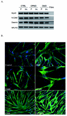
          Investigations into both the pathophysiology and therapeutic targets in muscle dystrophies have been hampered by the limited proliferative capacity of human myoblasts. Isolation of reliable and stable immortalized cell lines from patient biopsies is a powerful tool for investigating pathological mechanisms, including those associated with muscle aging, and for developing innovative gene-based, cell-based or pharmacological biotherapies.

          Methods

          Using transduction with both telomerase-expressing and cyclin-dependent kinase 4-expressing vectors, we were able to generate a battery of immortalized human muscle stem-cell lines from patients with various neuromuscular disorders.

          Results

          The immortalized human cell lines from patients with Duchenne muscular dystrophy, facioscapulohumeral muscular dystrophy, oculopharyngeal muscular dystrophy, congenital muscular dystrophy, and limb-girdle muscular dystrophy type 2B had greatly increased proliferative capacity, and maintained their potential to differentiate both in vitro and in vivo after transplantation into regenerating muscle of immunodeficient mice.

          Conclusions

          Dystrophic cellular models are required as a supplement to animal models to assess cellular mechanisms, such as signaling defects, or to perform high-throughput screening for therapeutic molecules. These investigations have been conducted for many years on cells derived from animals, and would greatly benefit from having human cell models with prolonged proliferative capacity. Furthermore, the possibility to assess in vivo the regenerative capacity of these cells extends their potential use. The innovative cellular tools derived from several different neuromuscular diseases as described in this report will allow investigation of the pathophysiology of these disorders and assessment of new therapeutic strategies.

          Related collections

          Most cited references35

          • Record: found
          • Abstract: found
          • Article: not found

          Extension of life-span by introduction of telomerase into normal human cells.

          Normal human cells undergo a finite number of cell divisions and ultimately enter a nondividing state called replicative senescence. It has been proposed that telomere shortening is the molecular clock that triggers senescence. To test this hypothesis, two telomerase-negative normal human cell types, retinal pigment epithelial cells and foreskin fibroblasts, were transfected with vectors encoding the human telomerase catalytic subunit. In contrast to telomerase-negative control clones, which exhibited telomere shortening and senescence, telomerase-expressing clones had elongated telomeres, divided vigorously, and showed reduced straining for beta-galactosidase, a biomarker for senescence. Notably, the telomerase-expressing clones have a normal karyotype and have already exceeded their normal life-span by at least 20 doublings, thus establishing a causal relationship between telomere shortening and in vitro cellular senescence. The ability to maintain normal human cells in a phenotypically youthful state could have important applications in research and medicine.
            Bookmark
            • Record: found
            • Abstract: found
            • Article: not found

            Telomere measurement by quantitative PCR.

            R. Cawthon (2002)
            It has long been presumed impossible to measure telomeres in vertebrate DNA by PCR amplification with oligonucleotide primers designed to hybridize to the TTAGGG and CCCTAA repeats, because only primer dimer-derived products are expected. Here we present a primer pair that eliminates this problem, allowing simple and rapid measurement of telomeres in a closed tube, fluorescence-based assay. This assay will facilitate investigations of the biology of telomeres and the roles they play in the molecular pathophysiology of diseases and aging.
              Bookmark
              • Record: found
              • Abstract: found
              • Article: found
              Is Open Access

              Telomere length measurement by a novel monochrome multiplex quantitative PCR method

              The current quantitative polymerase chain reaction (QPCR) assay of telomere length measures telomere (T) signals in experimental DNA samples in one set of reaction wells, and single copy gene (S) signals in separate wells, in comparison to a reference DNA, to yield relative T/S ratios that are proportional to average telomere length. Multiplexing this assay is desirable, because variation in the amount of DNA pipetted would no longer contribute to variation in T/S, since T and S would be collected within each reaction, from the same input DNA. Multiplexing also increases throughput and lowers costs, since half as many reactions are needed. Here, we present the first multiplexed QPCR method for telomere length measurement. Remarkably, a single fluorescent DNA-intercalating dye is sufficient in this system, because T signals can be collected in early cycles, before S signals rise above baseline, and S signals can be collected at a temperature that fully melts the telomere product, sending its signal to baseline. The correlation of T/S ratios with Terminal Restriction Fragment (TRF) lengths measured by Southern blot was stronger with this monochrome multiplex QPCR method (R 2 = 0.844) than with our original singleplex method (R 2 = 0.677). Multiplex T/S results from independent runs on different days were highly reproducible (R 2 = 0.91).
                Bookmark

                Author and article information

                Journal
                Skelet Muscle
                Skeletal Muscle
                BioMed Central
                2044-5040
                2011
                1 November 2011
                : 1
                : 34
                Affiliations
                [1 ]Thérapie des maladies du muscle strié, Institut de Myologie, UM76, UPMC Université Paris 6, Paris, France
                [2 ]INSERM U974, Paris, France
                [3 ]CNRS UMR 7215, Paris, France
                [4 ]Innate Immunity Unit, INSERM U 668, Institut Pasteur, Paris, France
                [5 ]Service d'Oto-Rhino-Laryngologie et de Chirurgie Cervico-Faciale, Faculté de Médecine St Antoine, Université Pierre et Marie Curie, Hôpital Tenon, Paris, France
                [6 ]The Dubowitz Neuromuscular Centre, Institute of Child Health, University College, London, UK
                [7 ]Muscle Research Unit, Experimental and Clinical Research Center, Charité University Hospital and Max Delbrück Center for Molecular Medicine, Berlin, Germany
                [8 ]Faculté de Médecine de Marseille, Université de la Méditerranée, Inserm UMRS 910 Génétique Médicale et Génomique Fonctionnelle, Marseille, France
                [9 ]Department of Neurology, Hillel Yaffe Medical Center, PO Box 169, Hadera, 38100, Israel
                [10 ]UT Southwestern Medical Center, Department of Cell Biology, Dallas, TX 75390, USA
                [11 ]Laboratoire LBCM, Departement de Biologie, Faculté des Sciences, Agadir, Maroc
                Article
                2044-5040-1-34
                10.1186/2044-5040-1-34
                3235972
                22040608
                6212d7a1-0aa6-4c6e-b702-d3ed3251be83
                Copyright ©2011 Mamchaoui et al; licensee BioMed Central Ltd.

                This is an Open Access article distributed under the terms of the Creative Commons Attribution License ( http://creativecommons.org/licenses/by/2.0), which permits unrestricted use, distribution, and reproduction in any medium, provided the original work is properly cited.

                History
                : 20 June 2011
                : 1 November 2011
                Categories
                Research

                Rheumatology
                Rheumatology

                Comments

                Comment on this article