16
views
0
recommends
+1 Recommend
0 collections
    0
    shares
      • Record: found
      • Abstract: found
      • Article: found
      Is Open Access

      Development of a Three‐Dimensional Bioengineering Technology to Generate Lung Tissue for Personalized Disease Modeling

      research-article

      Read this article at

      Bookmark
          There is no author summary for this article yet. Authors can add summaries to their articles on ScienceOpen to make them more accessible to a non-specialist audience.

          Abstract

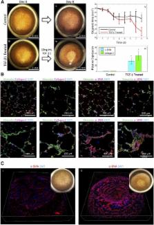
          Stem cell technologies, especially patient‐specific, induced stem cell pluripotency and directed differentiation, hold great promise for changing the landscape of medical therapies. Proper exploitation of these methods may lead to personalized organ transplants, but to regenerate organs, it is necessary to develop methods for assembling differentiated cells into functional, organ‐level tissues. The generation of three‐dimensional human tissue models also holds potential for medical advances in disease modeling, as full organ functionality may not be necessary to recapitulate disease pathophysiology. This is specifically true of lung diseases where animal models often do not recapitulate human disease. Here, we present a method for the generation of self‐assembled human lung tissue and its potential for disease modeling and drug discovery for lung diseases characterized by progressive and irreversible scarring such as idiopathic pulmonary fibrosis (IPF). Tissue formation occurs because of the overlapping processes of cellular adhesion to multiple alveolar sac templates, bioreactor rotation, and cellular contraction. Addition of transforming growth factor‐β1 to single cell‐type mesenchymal organoids resulted in morphologic scarring typical of that seen in IPF but not in two‐dimensional IPF fibroblast cultures. Furthermore, this lung organoid may be modified to contain multiple lung cell types assembled into the correct anatomical location, thereby allowing cell‐cell contact and recapitulating the lung microenvironment. Our bottom‐up approach for synthesizing patient‐specific lung tissue in a scalable system allows for the development of relevant human lung disease models with the potential for high throughput drug screening to identify targeted therapies. S tem C ells T ranslational M edicine 2017;6:622–633

          Related collections

          Most cited references29

          • Record: found
          • Abstract: found
          • Article: not found

          Idiopathic pulmonary fibrosis: prevailing and evolving hypotheses about its pathogenesis and implications for therapy.

          Idiopathic pulmonary fibrosis is a progressive and usually fatal lung disease characterized by fibroblast proliferation and extracellular matrix remodeling, which result in irreversible distortion of the lung's architecture. Although the pathogenetic mechanisms remain to be determined, the prevailing hypothesis holds that fibrosis is preceded and provoked by a chronic inflammatory process that injures the lung and modulates lung fibrogenesis, leading to the end-stage fibrotic scar. However, there is little evidence that inflammation is prominent in early disease, and it is unclear whether inflammation is relevant to the development of the fibrotic process. Evidence suggests that inflammation does not play a pivotal role. Inflammation is not a prominent histopathologic finding, and epithelial injury in the absence of ongoing inflammation is sufficient to stimulate the development of fibrosis. In addition, the inflammatory response to a lung fibrogenic insult is not necessarily related to the fibrotic response. Clinical measurements of inflammation fail to correlate with stage or outcome, and potent anti-inflammatory therapy does not improve outcome. This review presents a growing body of evidence suggesting that idiopathic pulmonary fibrosis involves abnormal wound healing in response to multiple, microscopic sites of ongoing alveolar epithelial injury and activation associated with the formation of patchy fibroblast-myofibroblast foci, which evolve to fibrosis. Progress in understanding the fibrogenic mechanisms in the lung is likely to yield more effective therapies.
            Bookmark
            • Record: found
            • Abstract: found
            • Article: not found

            Lung stem cell differentiation in mice directed by endothelial cells via a BMP4-NFATc1-thrombospondin-1 axis.

            Lung stem cells are instructed to produce lineage-specific progeny through unknown factors in their microenvironment. We used clonal 3D cocultures of endothelial cells and distal lung stem cells, bronchioalveolar stem cells (BASCs), to probe the instructive mechanisms. Single BASCs had bronchiolar and alveolar differentiation potential in lung endothelial cell cocultures. Gain- and loss-of-function experiments showed that BMP4-Bmpr1a signaling triggers calcineurin/NFATc1-dependent expression of thrombospondin-1 (Tsp1) in lung endothelial cells to drive alveolar lineage-specific BASC differentiation. Tsp1 null mice exhibited defective alveolar injury repair, confirming a crucial role for the BMP4-NFATc1-TSP1 axis in lung epithelial differentiation and regeneration in vivo. Discovery of this pathway points to methods to direct the derivation of specific lung epithelial lineages from multipotent cells. These findings elucidate a pathway that may be a critical target in lung diseases and provide tools to understand the mechanisms of respiratory diseases at the single-cell level. Copyright © 2014 Elsevier Inc. All rights reserved.
              Bookmark
              • Record: found
              • Abstract: found
              • Article: not found

              Vascularized and Complex Organ Buds from Diverse Tissues via Mesenchymal Cell-Driven Condensation.

              Transplantation of in-vitro-generated organ buds is a promising approach toward regenerating functional and vascularized organs. Though it has been recently shown in the context of liver models, demonstrating the applicability of this approach to other systems by delineating the molecular mechanisms guiding organ bud formation is critical. Here, we demonstrate a generalized method for organ bud formation from diverse tissues by combining pluripotent stem cell-derived tissue-specific progenitors or relevant tissue samples with endothelial cells and mesenchymal stem cells (MSCs). The MSCs initiated condensation within these heterotypic cell mixtures, which was dependent upon substrate matrix stiffness. Defining optimal mechanical properties promoted formation of 3D, transplantable organ buds from tissues including kidney, pancreas, intestine, heart, lung, and brain. Transplanted pancreatic and renal buds were rapidly vascularized and self-organized into functional, tissue-specific structures. These findings provide a general platform for harnessing mechanical properties to generate vascularized, complex organ buds with broad applications for regenerative medicine.
                Bookmark

                Author and article information

                Contributors
                bgomperts@mednet.ucla.edu
                Journal
                Stem Cells Transl Med
                Stem Cells Transl Med
                10.1002/(ISSN)2157-6580
                SCT3
                Stem Cells Translational Medicine
                John Wiley and Sons Inc. (Hoboken )
                2157-6564
                2157-6580
                15 September 2016
                February 2017
                : 6
                : 2 ( doiID: 10.1002/sct3.2017.6.issue-2 )
                : 622-633
                Affiliations
                [ 1 ]Department of Materials Science and Engineering, University of California, Los Angeles, Los Angeles, California, USA
                [ 2 ]David Geffen School of Medicine at the University of California, Los Angeles, Department of Pediatrics, Children's Discovery and Innovation Institute, Los Angeles, California, USA
                [ 3 ]Eli and Edythe Broad Center of Regenerative Medicine and Stem Cell Research, University of California, Los Angeles, Los Angeles, California, USA
                Author notes
                [*] [* ]Correspondence: Brigitte N. Gomperts, M.D., David Geffen School of Medicine, Department of Pediatrics, University of California, Los Angeles, Los Angeles, California 90095, USA. Telephone: 310‐206‐0711; e‐mail: bgomperts@ 123456mednet.ucla.edu
                Article
                SCT312094
                10.5966/sctm.2016-0192
                5442826
                28191779
                62f10a79-443c-4c68-8bbe-eeb640fef510
                © 2016 The Authors S tem C ells T ranslational M edicine published by Wiley Periodicals, Inc. on behalf of AlphaMed Press

                This is an open access article under the terms of the Creative Commons Attribution License, which permits use, distribution and reproduction in any medium, provided the original work is properly cited.

                History
                : 14 April 2016
                : 01 August 2016
                Page count
                Figures: 5, Tables: 0, Pages: 12, Words: 8749
                Categories
                Tissue Engineering and Regenerative Medicine
                Translational Research Articles and Reviews
                Tissue Engineering and Regenerative Medicine
                Custom metadata
                2.0
                sct312094
                February 2017
                Converter:WILEY_ML3GV2_TO_NLMPMC version:5.0.9 mode:remove_FC converted:23.05.2017

                lung,tissue engineering,disease modeling,three‐dimensional cell culture

                Comments

                Comment on this article