12
views
0
recommends
+1 Recommend
0 collections
    0
    shares
      • Record: found
      • Abstract: found
      • Article: found
      Is Open Access

      Patho- physiological role of BDNF in fibrin clotting

      research-article

      Read this article at

      Bookmark
          There is no author summary for this article yet. Authors can add summaries to their articles on ScienceOpen to make them more accessible to a non-specialist audience.

          Abstract

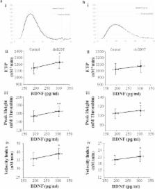
          Circulating levels of Brain Derived Neurotrophic Factor (BDNF) are lower in coronary heart disease (CHD) than in healthy subjects and are associated with coronary events and mortality. However, the mechanism(s) underling this association is not fully understood. We hypothesize that BDNF may influence fibrin fiber structure and clot stability, favoring clot lysis and thrombus resolution. We showed that recombinant BDNF (rh-BDNF) influenced with clot formation in a concentration-dependent manner in both purified fibrinogen and plasma from healthy subjects. In particular, rh-BDNF reduced the density of fibrin fibers, the maximum clot firmness (MCF) and the maximum clot turbidity, and affected the lysis of clot. In addition, both thrombin and reptilase clotting time were prolonged by rh-BDNF, despite the amount of thrombin formed was greater. Intriguingly, CHD patients had lower levels of BDNF, greater fibrin fibers density, higher MCF than control subjects, and a negative correlation between BDNF and MCF was found. Of note, rh-BDNF markedly modified fibrin clot profile restoring physiological clot morphology in CHD plasma. In conclusion, we provide evidence that low levels of BDNF correlate with the formation of bigger thrombi ( in vitro) and that this effect is mediated, at least partially, by the alteration of fibrin fibers formation.

          Related collections

          Most cited references49

          • Record: found
          • Abstract: found
          • Article: not found

          Heparin-binding domain of fibrin(ogen) binds growth factors and promotes tissue repair when incorporated within a synthetic matrix.

          By binding growth factors (GFs), the ECM tightly regulates their activity. We recently reported that the heparin-binding domain II of fibronectin acts as a promiscuous high-affinity GF-binding domain. Here we hypothesized that fibrin, the provisional ECM during tissue repair, also could be highly promiscuous in its GF-binding capacity. Using multiple affinity-based assays, we found that fibrin(ogen) and its heparin-binding domain bind several GFs from the PDGF/VEGF and FGF families and some GFs from the TGF-β and neurotrophin families. Overall, we identified 15 unique binding interactions. The GF binding ability of fibrinogen caused prolonged retention of many of the identified GFs within fibrin. Thus, based on the promiscuous and high-affinity interactions in fibrin, GF binding may be one of fibrin's main physiological functions, and these interactions may potentially play an important and ubiquitous role during tissue repair. To prove this role in a gain-of-function model, we incorporated the heparin-binding domain of fibrin into a synthetic fibrin-mimetic matrix. In vivo, the multifunctional synthetic matrix could fully mimic the effect of fibrin in a diabetic mouse model of impaired wound healing, demonstrating the benefits of generating a hybrid biomaterial consisting of a synthetic polymeric scaffold and recombinant bioactive ECM domains. The reproduction of GF-ECM interactions with a fibrin-mimetic matrix could be clinically useful, and has the significant benefit of a more straightforward regulatory path associated with chemical synthesis rather than human sourcing.
            Bookmark
            • Record: found
            • Abstract: found
            • Article: not found

            Purification of a new neurotrophic factor from mammalian brain.

            We report the purification from pig brain of a factor supporting the survival of, and fibre outgrowth from, cultured embryonic chick sensory neurons. The purified factor migrates as one single band, mol. wt. 12 300, on gel electrophoresis in the presence of sodium dodecylsulphate (SDS) and is a basic molecule (pI greater than or equal to 10.1). Approximately 1 microgram factor was isolated from 1.5 kg brain. The final degree of purification was estimated to be 1.4 X 10(6)-fold, and the specific activity 0.4 ng/ml/unit, which is similar to that of nerve growth factor (NGF) using the same assay system. This factor is the first neurotrophic factor to be purified since NGF, from which it is clearly distinguished because it has different antigenic and functional properties.
              Bookmark
              • Record: found
              • Abstract: not found
              • Article: not found

              A convenient method for quantifying collagen fibers in atherosclerotic lesions by ImageJ software

                Bookmark

                Author and article information

                Contributors
                silvia.barbieri@ccfm.it
                Journal
                Sci Rep
                Sci Rep
                Scientific Reports
                Nature Publishing Group UK (London )
                2045-2322
                23 January 2019
                23 January 2019
                2019
                : 9
                : 389
                Affiliations
                [1 ]GRID grid.414603.4, Centro Cardiologico Monzino, , IRCCS, ; Milan, Italy
                [2 ]ISNI 0000 0004 1757 2822, GRID grid.4708.b, Dipartimento di Scienze Farmacologiche e Biomolecolari, , Università degli Studi di Milano, ; Milan, Italy
                Author information
                http://orcid.org/0000-0002-4019-4618
                Article
                37117
                10.1038/s41598-018-37117-1
                6344484
                30674980
                639045d1-ddba-4bf2-a68c-e729f5118275
                © The Author(s) 2019

                Open Access This article is licensed under a Creative Commons Attribution 4.0 International License, which permits use, sharing, adaptation, distribution and reproduction in any medium or format, as long as you give appropriate credit to the original author(s) and the source, provide a link to the Creative Commons license, and indicate if changes were made. The images or other third party material in this article are included in the article’s Creative Commons license, unless indicated otherwise in a credit line to the material. If material is not included in the article’s Creative Commons license and your intended use is not permitted by statutory regulation or exceeds the permitted use, you will need to obtain permission directly from the copyright holder. To view a copy of this license, visit http://creativecommons.org/licenses/by/4.0/.

                History
                : 25 July 2018
                : 3 December 2018
                Funding
                Funded by: Sandrini L is supported by the 32nd cycle PhD program in Scienze Farmacologiche Sperimentali e Cliniche, Università degli Studi di Milano.
                Funded by: FundRef https://doi.org/10.13039/501100003197, Ministry of Health, Italy | Agenzia Italiana del Farmaco, Ministero della Salute (Italian Medicines Agency);
                Award ID: RC: BIO37-2016: 2613074; BIO37-2017: 2631213
                Award Recipient :
                Funded by: Italian Ministry of Health: Italian “5 × 1000” tax (2014 and 2015)
                Categories
                Article
                Custom metadata
                © The Author(s) 2019

                Uncategorized
                Uncategorized

                Comments

                Comment on this article