95
views
0
recommends
+1 Recommend
0 collections
    0
    shares
      • Record: found
      • Abstract: found
      • Article: found
      Is Open Access

      Safety and Efficacy of Ranibizumab in Diabetic Macular Edema (RESOLVE Study*) : A 12-month, randomized, controlled, double-masked, multicenter phase II study

      research-article

      Read this article at

      Bookmark
          There is no author summary for this article yet. Authors can add summaries to their articles on ScienceOpen to make them more accessible to a non-specialist audience.

          Abstract

          OBJECTIVE

          The expression of vascular endothelial growth factor (VEGF) is elevated in diabetic macular edema (DME). Ranibizumab binds to and inhibits multiple VEGF variants. We investigated the safety and efficacy of ranibizumab in DME involving the foveal center.

          RESEARCH DESIGN AND METHODS

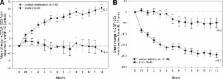
          This was a 12-month, multicenter, sham-controlled, double-masked study with eyes (age >18 years, type 1 or 2 diabetes, central retinal thickness [CRT] ≥300 μm, and best corrected visual acuity [BCVA] of 73–39 ETDRS letters [Early Treatment Diabetic Retinopathy Study]) randomly assigned to intravitreal ranibizumab (0.3 or 0.5 mg; n = 51 each) or sham ( n = 49). The treatment schedule comprised three monthly injections, after which treatment could be stopped/reinitiated with an opportunity for rescue laser photocoagulation (protocol-defined criteria). After month 1, dose-doubling was permitted (protocol-defined criteria, injection volume increased from 0.05 to 0.1 ml and remained at 0.1 ml thereafter). Efficacy (BCVA and CRT) and safety were compared between pooled ranibizumab and sham arms using the full analysis set ( n = 151, patients receiving ≥1 injection).

          RESULTS

          At month 12, mean ± SD BCVA improved from baseline by 10.3 ± 9.1 letters with ranibizumab and declined by 1.4 ± 14.2 letters with sham ( P < 0.0001). Mean CRT reduction was 194.2 ± 135.1 μm with ranibizumab and 48.4 ± 153.4 μm with sham ( P < 0.0001). Gain of ≥10 letters BCVA from baseline occurred in 60.8% of ranibizumab and 18.4% of sham eyes ( P < 0.0001). Safety data were consistent with previous studies of intravitreal ranibizumab.

          CONCLUSIONS

          Ranibizumab is effective in improving BCVA and is well tolerated in DME. Future clinical trials are required to confirm its long-term efficacy and safety.

          Related collections

          Most cited references16

          • Record: found
          • Abstract: found
          • Article: not found

          Global data on visual impairment in the year 2002.

          This paper presents estimates of the prevalence of visual impairment and its causes in 2002, based on the best available evidence derived from recent studies. Estimates were determined from data on low vision and blindness as defined in the International statistical classification of diseases, injuries and causes of death, 10th revision. The number of people with visual impairment worldwide in 2002 was in excess of 161 million, of whom about 37 million were blind. The burden of visual impairment is not distributed uniformly throughout the world: the least developed regions carry the largest share. Visual impairment is also unequally distributed across age groups, being largely confined to adults 50 years of age and older. A distribution imbalance is also found with regard to gender throughout the world: females have a significantly higher risk of having visual impairment than males. Notwithstanding the progress in surgical intervention that has been made in many countries over the last few decades, cataract remains the leading cause of visual impairment in all regions of the world, except in the most developed countries. Other major causes of visual impairment are, in order of importance, glaucoma, age-related macular degeneration, diabetic retinopathy and trachoma.
            Bookmark
            • Record: found
            • Abstract: found
            • Article: not found

            Randomized trial evaluating ranibizumab plus prompt or deferred laser or triamcinolone plus prompt laser for diabetic macular edema.

            Evaluate intravitreal 0.5 mg ranibizumab or 4 mg triamcinolone combined with focal/grid laser compared with focal/grid laser alone for treatment of diabetic macular edema (DME). Multicenter, randomized clinical trial. A total of 854 study eyes of 691 participants with visual acuity (approximate Snellen equivalent) of 20/32 to 20/320 and DME involving the fovea. Eyes were randomized to sham injection + prompt laser (n=293), 0.5 mg ranibizumab + prompt laser (n=187), 0.5 mg ranibizumab + deferred (> or =24 weeks) laser (n=188), or 4 mg triamcinolone + prompt laser (n=186). Retreatment followed an algorithm facilitated by a web-based, real-time data-entry system. Best-corrected visual acuity and safety at 1 year. The 1-year mean change (+/-standard deviation) in the visual acuity letter score from baseline was significantly greater in the ranibizumab + prompt laser group (+9+/-11, P<0.001) and ranibizumab + deferred laser group (+9+/-12, P<0.001) but not in the triamcinolone + prompt laser group (+4+/-13, P=0.31) compared with the sham + prompt laser group (+3+/-13). Reduction in mean central subfield thickness in the triamcinolone + prompt laser group was similar to both ranibizumab groups and greater than in the sham + prompt laser group. In the subset of pseudophakic eyes at baseline (n=273), visual acuity improvement in the triamcinolone + prompt laser group appeared comparable to that in the ranibizumab groups. No systemic events attributable to study treatment were apparent. Three eyes (0.8%) had injection-related endophthalmitis in the ranibizumab groups, whereas elevated intraocular pressure and cataract surgery were more frequent in the triamcinolone + prompt laser group. Two-year visual acuity outcomes were similar to 1-year outcomes. Intravitreal ranibizumab with prompt or deferred laser is more effective through at least 1 year compared with prompt laser alone for the treatment of DME involving the central macula. Ranibizumab as applied in this study, although uncommonly associated with endophthalmitis, should be considered for patients with DME and characteristics similar to those in this clinical trial. In pseudophakic eyes, intravitreal triamcinolone + prompt laser seems more effective than laser alone but frequently increases the risk of intraocular pressure elevation. Copyright 2010 American Academy of Ophthalmology. Published by Elsevier Inc. All rights reserved.
              Bookmark
              • Record: found
              • Abstract: found
              • Article: not found

              Photocoagulation for diabetic macular edema. Early Treatment Diabetic Retinopathy Study report number 1. Early Treatment Diabetic Retinopathy Study research group.

              (1985)
              Data from the Early Treatment Diabetic Retinopathy Study (ETDRS) show that focal photocoagulation of "clinically significant" diabetic macular edema substantially reduces the risk of visual loss. Focal treatment also increases the chance of visual improvement, decreases the frequency of persistent macular edema, and causes only minor visual field losses. In this randomized clinical trial, which was supported by the National Eye Institute, 754 eyes that had macular edema and mild to moderate diabetic retinopathy were randomly assigned to focal argon laser photocoagulation, while 1,490 such eyes were randomly assigned to deferral of photocoagulation. The beneficial effects of treatment demonstrated in this trial suggest that all eyes with clinically significant diabetic macular edema should be considered for focal photocoagulation. Clinically significant macular edema is defined as retinal thickening that involves or threatens the center of the macula (even if visual acuity is not yet reduced) and is assessed by stereo contact lens biomicroscopy or stereo photography. Follow-up of all ETDRS patients continues without other modifications in the study protocol.
                Bookmark

                Author and article information

                Journal
                Diabetes Care
                diacare
                dcare
                Diabetes Care
                Diabetes Care
                American Diabetes Association
                0149-5992
                1935-5548
                November 2010
                : 33
                : 11
                : 2399-2405
                Affiliations
                [1] 1Assistance Publique des Hôpitaux de Paris, Ophthalmology Department, Hôpital Lariboisière, Paris, France;
                [2] 2Department of Ophthalmology, University of Udine, Udine, Italy;
                [3] 3Clinic for Vitreoretinal Disease, Swiss Eye Institute and University of Bern, Bern, Switzerland;
                [4] 4University Hospital Freiburg, Freiburg, Germany;
                [5] 5Ophthalmology Research Unit, School of Clinical Sciences, University of Liverpool, Liverpool, U.K.;
                [6] 6Department of Ophthalmology, Glostrup Hospital, University of Copenhagen, Copenhagen, Denmark;
                [7] 7Centre for Vision Research, University of Sydney, Sydney, New South Wales, Australia;
                [8] 8Department of Ophthalmology, University of Auckland, Auckland, New Zealand;
                [9] 9Bern Photographic Reading Center, Department of Ophthalmology, Inselspital, University Bern, Bern, Switzerland;
                [10] 10Novartis Pharma, Basel, Switzerland;
                [11] 11Department of Ophthalmology, Inselspital, University Bern, Bern, Switzerland.
                Author notes
                Corresponding author: Sebastian Wolf, sebastian.wolf@ 123456insel.ch .

                F.B. is currently affiliated with the Department of Ophthalmology, University Vita Salute, Scientific Institute San Raffaele, Milan, Italy.

                *A complete list of the RESOLVE investigators can be found in the online appendix, available at http://care.diabetesjournals.org/cgi/content/full/dc10-0493/DC1.

                Article
                0493
                10.2337/dc10-0493
                2963502
                20980427
                648edfcf-6514-4bab-8199-b85f1d885151
                © 2010 by the American Diabetes Association.

                Readers may use this article as long as the work is properly cited, the use is educational and not for profit, and the work is not altered. See http://creativecommons.org/licenses/by-nc-nd/3.0/ for details.

                History
                : 15 March 2010
                : 8 August 2010
                Categories
                Original Research
                Emerging Treatments and Technologies

                Endocrinology & Diabetes
                Endocrinology & Diabetes

                Comments

                Comment on this article