2
views
0
recommends
+1 Recommend
0 collections
    0
    shares
      • Record: found
      • Abstract: found
      • Article: found
      Is Open Access

      Higher testosterone and testosterone/estradiol ratio in men are associated with better epigenetic estimators of mortality risk

      Preprint
      research-article

      Read this article at

      Bookmark
          There is no author summary for this article yet. Authors can add summaries to their articles on ScienceOpen to make them more accessible to a non-specialist audience.

          Abstract

          Introduction:

          Sex hormones are hypothesized to drive sex-specific health disparities. Here, we study the association between sex steroid hormones and DNA methylation-based (DNAm) biomarkers of age and mortality risk including Pheno Age Acceleration (AA), Grim AA, and DNAm-based estimators of Plasminogen Activator Inhibitor 1 (PAI1), and leptin concentrations.

          Methods:

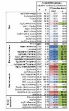
          We pooled data from three population-based cohorts, the Framingham Heart Study Offspring Cohort (FHS), the Baltimore Longitudinal Study of Aging (BLSA), and the InCHIANTI Study, including 1,062 postmenopausal women without hormone therapy and 1,612 men of European descent. Sex hormone concentrations were standardized with mean 0 and standard deviation of 1, for each study and sex separately. Sex-stratified analyses using a linear mixed regression were performed, with a Benjamini-Hochberg (BH) adjustment for multiple testing. Sensitivity analysis was performed excluding the previously used training-set for the development of Pheno and Grim age.

          Results:

          Sex Hormone Binding Globulin (SHBG) is associated with a decrease in DNAm PAI1 among men (per 1 standard deviation (SD): −478 pg/mL; 95%CI: −614 to −343; P:1e-11; BH-P: 1e-10), and women (−434 pg/mL; 95%CI: −589 to −279; P:1e-7; BH-P:2e-6). The testosterone/estradiol (TE) ratio was associated with a decrease in Pheno AA (−0.41 years; 95%CI: −0.70 to −0.12; P:0.01; BH-P: 0.04), and DNAm PAI1 (−351 pg/mL; 95%CI: −486 to −217; P:4e-7; BH-P:3e-6) among men. In men, 1 SD increase in total testosterone was associated with a decrease in DNAm PAI1 (−481 pg/mL; 95%CI: −613 to −349; P:2e-12; BH-P:6e-11).

          Conclusion:

          SHBG was associated with lower DNAm PAI1 among men and women. Higher testosterone and testosterone/estradiol ratio were associated with lower DNAm PAI and a younger epigenetic age in men. A decrease in DNAm PAI1 is associated with lower mortality and morbidity risk indicating a potential protective effect of testosterone on lifespan and conceivably cardiovascular health via DNAm PAI1.

          Visual representation of our main results stratified by sex.

          There were four outcomes of interest in the rectangular shapes in the middle of this figure, Pheno-Age Acceleration (AA), Grim AA, DNAm-based PAI1, and DNAm-based leptin. We measured five hormone concentrations (testosterone, estrone, estradiol, DHEAS, and Sex Hormone Binding Globulin (SHBG)). In addition, one hormone level ratio (testosterone / estradiol) was estimated. Associations were calculated by linear mixed regression models between sex hormones and the outcomes of interests. The associations are represented by colored arrows with the lines’ thickness representing the strength of the association. As the association was measured mainly cross-sectional, the directionality of the association cannot be established. Hormone levels were inversely associated with epigenetic estimators of mortality risk.

          Abbreviations: E1: total estrone; E2: total estradiol; SHBG: Sex Hormone Binding Globulin; TotT: total testosterone; TE ratio: Total testosterone divided by total estradiol concentration

          Related collections

          Most cited references96

          • Record: found
          • Abstract: found
          • Article: not found

          DNA methylation age of human tissues and cell types

          Background It is not yet known whether DNA methylation levels can be used to accurately predict age across a broad spectrum of human tissues and cell types, nor whether the resulting age prediction is a biologically meaningful measure. Results I developed a multi-tissue predictor of age that allows one to estimate the DNA methylation age of most tissues and cell types. The predictor, which is freely available, was developed using 8,000 samples from 82 Illumina DNA methylation array datasets, encompassing 51 healthy tissues and cell types. I found that DNA methylation age has the following properties: first, it is close to zero for embryonic and induced pluripotent stem cells; second, it correlates with cell passage number; third, it gives rise to a highly heritable measure of age acceleration; and, fourth, it is applicable to chimpanzee tissues. Analysis of 6,000 cancer samples from 32 datasets showed that all of the considered 20 cancer types exhibit significant age acceleration, with an average of 36 years. Low age-acceleration of cancer tissue is associated with a high number of somatic mutations and TP53 mutations, while mutations in steroid receptors greatly accelerate DNA methylation age in breast cancer. Finally, I characterize the 353 CpG sites that together form an aging clock in terms of chromatin states and tissue variance. Conclusions I propose that DNA methylation age measures the cumulative effect of an epigenetic maintenance system. This novel epigenetic clock can be used to address a host of questions in developmental biology, cancer and aging research.
            Bookmark
            • Record: found
            • Abstract: found
            • Article: found
            Is Open Access

            An epigenetic biomarker of aging for lifespan and healthspan

            Identifying reliable biomarkers of aging is a major goal in geroscience. While the first generation of epigenetic biomarkers of aging were developed using chronological age as a surrogate for biological age, we hypothesized that incorporation of composite clinical measures of phenotypic age that capture differences in lifespan and healthspan may identify novel CpGs and facilitate the development of a more powerful epigenetic biomarker of aging. Using an innovative two-step process, we develop a new epigenetic biomarker of aging, DNAm PhenoAge, that strongly outperforms previous measures in regards to predictions for a variety of aging outcomes, including all-cause mortality, cancers, healthspan, physical functioning, and Alzheimer's disease. While this biomarker was developed using data from whole blood, it correlates strongly with age in every tissue and cell tested. Based on an in-depth transcriptional analysis in sorted cells, we find that increased epigenetic, relative to chronological age, is associated with increased activation of pro-inflammatory and interferon pathways, and decreased activation of transcriptional/translational machinery, DNA damage response, and mitochondrial signatures. Overall, this single epigenetic biomarker of aging is able to capture risks for an array of diverse outcomes across multiple tissues and cells, and provide insight into important pathways in aging.
              Bookmark
              • Record: found
              • Abstract: found
              • Article: not found

              Genome-wide methylation profiles reveal quantitative views of human aging rates.

              The ability to measure human aging from molecular profiles has practical implications in many fields, including disease prevention and treatment, forensics, and extension of life. Although chronological age has been linked to changes in DNA methylation, the methylome has not yet been used to measure and compare human aging rates. Here, we build a quantitative model of aging using measurements at more than 450,000 CpG markers from the whole blood of 656 human individuals, aged 19 to 101. This model measures the rate at which an individual's methylome ages, which we show is impacted by gender and genetic variants. We also show that differences in aging rates help explain epigenetic drift and are reflected in the transcriptome. Moreover, we show how our aging model is upheld in other human tissues and reveals an advanced aging rate in tumor tissue. Our model highlights specific components of the aging process and provides a quantitative readout for studying the role of methylation in age-related disease. Copyright © 2013 Elsevier Inc. All rights reserved.
                Bookmark

                Author and article information

                Journal
                medRxiv
                MEDRXIV
                medRxiv
                Cold Spring Harbor Laboratory
                30 July 2023
                : 2023.02.16.23285997
                Affiliations
                [1 ]Department of Human Genetics, David Geffen School of Medicine, Los Angeles, CA, USA
                [2 ]Department of Neurology, David Geffen School of Medicine, Los Angeles, CA, USA.
                [3 ]Altos Labs, San Diego, USA
                [4 ]Longitudinal Studies Section, Translational Gerontology Branch, National Institute on Aging, National Institutes of Health, Baltimore, USA
                [5 ]Department of Epidemiology, UCLA Fielding School of Public Health, Los Angeles, CA, USA
                [6 ]Department of Environmental Health, UCLA Fielding School of Public Health, Los Angeles, CA, USA.
                [7 ]Population Sciences in the Pacific Program, University of Hawaii Cancer Center, Honolulu, Hawaii, USA
                [8 ]Department of Biostatistics, School of Public Health, University of California, Los Angeles, Los Angeles, CA, USA
                Author notes
                Corresponding author: Cynthia DJ Kusters, Present address: Department of Epidemiology, Fielding School of Public Health at UCLA, 650 Charles E. Young Drive South, Box 708822, Los Angeles, CA 90095-7088, USA. Tel: 310-422-2954. ckusters@ 123456ucla.edu
                Article
                10.1101/2023.02.16.23285997
                9980235
                36865294
                657981f6-7c0f-443e-8379-c76d5e5fe7da

                This work is licensed under a Creative Commons Attribution 4.0 International License, which allows reusers to distribute, remix, adapt, and build upon the material in any medium or format, so long as attribution is given to the creator. The license allows for commercial use.

                History
                Funding
                Funded by: NIH
                Award ID: F32AG063442
                Award ID: K01AG072044
                Award ID: U01AG060908
                Funded by: Intramural Research Program of the National Institute on Aging, NIH
                Funded by: The InCHIANTI study
                Award ID: ICS 110.1/RS97.71
                Funded by: Italian Ministry of Health, U.S. National Institute on Aging
                Award ID: N01-AG-916413
                Award ID: N01-AG-5-0002
                Award ID: N01-AG-821336
                Award ID: R01-AG-027012
                Funded by: National Cancer Institute of the National Institutes of Health
                Award ID: K07CA225856
                Funded by: National Institutes of Health
                Categories
                Article

                Comments

                Comment on this article