17
views
0
recommends
+1 Recommend
0 collections
    0
    shares
      • Record: found
      • Abstract: found
      • Article: found
      Is Open Access

      Modifications of Dental Implant Surfaces at the Micro- and Nano-Level for Enhanced Osseointegration

      review-article
      Materials
      MDPI
      surface modification, osseointegration, SLA, TiO2 nanotube, fluoride, photofunctionalization

      Read this article at

      Bookmark
          There is no author summary for this article yet. Authors can add summaries to their articles on ScienceOpen to make them more accessible to a non-specialist audience.

          Abstract

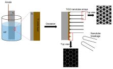
          This review paper describes several recent modification methods for biocompatible titanium dental implant surfaces. The micro-roughened surfaces reviewed in the literature are sandblasted, large-grit, acid-etched, and anodically oxidized. These globally-used surfaces have been clinically investigated, showing survival rates higher than 95%. In the past, dental clinicians believed that eukaryotic cells for osteogenesis did not recognize the changes of the nanostructures of dental implant surfaces. However, research findings have recently shown that osteogenic cells respond to chemical and morphological changes at a nanoscale on the surfaces, including titanium dioxide nanotube arrangements, functional peptide coatings, fluoride treatments, calcium–phosphorus applications, and ultraviolet photofunctionalization. Some of the nano-level modifications have not yet been clinically evaluated. However, these modified dental implant surfaces at the nanoscale have shown excellent in vitro and in vivo results, and thus promising potential future clinical use.

          Related collections

          Most cited references124

          • Record: found
          • Abstract: not found
          • Article: not found

          Light-induced amphiphilic surfaces

            Bookmark
            • Record: found
            • Abstract: found
            • Article: not found

            Stem cell fate dictated solely by altered nanotube dimension.

            Two important goals in stem cell research are to control the cell proliferation without differentiation and to direct the differentiation into a specific cell lineage when desired. Here, we demonstrate such paths by controlling only the nanotopography of culture substrates. Altering the dimensions of nanotubular-shaped titanium oxide surface structures independently allowed either augmented human mesenchymal stem cell (hMSC) adhesion or a specific differentiation of hMSCs into osteoblasts by using only the geometric cues, absent of osteogenic inducing media. hMSC behavior in response to defined nanotube sizes revealed a very dramatic change in hMSC behavior in a relatively narrow range of nanotube dimensions. Small (approximately 30-nm diameter) nanotubes promoted adhesion without noticeable differentiation, whereas larger (approximately 70- to 100-nm diameter) nanotubes elicited a dramatic stem cell elongation (approximately 10-fold increased), which induced cytoskeletal stress and selective differentiation into osteoblast-like cells, offering a promising nanotechnology-based route for unique orthopedics-related hMSC treatments.
              Bookmark
              • Record: found
              • Abstract: found
              • Article: not found

              Osseointegrated titanium implants. Requirements for ensuring a long-lasting, direct bone-to-implant anchorage in man.

              A total of 2895 threaded, cylindrical titanium implants have been inserted into the mandible or the maxilla and 124 similar implants have been installed in the tibial, temporal or iliac bones in man for various bone restorative procedures. The titanium screws were implanted without the use of cement, using a meticulous technique aiming at osseointegration--a direct contact between living bone and implant. Thirty-eight stable and integrated screws were removed for various reasons from 18 patients. The interface zone between bone and implant was investigated using X-rays, SEM, TEM and histology. The SEM study showed a very close spatial relationship between titanium and bone. The pattern of the anchorage of collagen filaments to titanium appeared to be similar to that of Sharpey's fibres to bone. No wear products were seen in the bone or soft tissues in spite of implant loading times up to 90 months. The soft tissues were also closely adhered to the titanium implant, thereby forming a biological seal, preventing microorganism infiltration along the implant. The implants in many cases had been allowed to permanently penetrate the gingiva and skin. This caused no adverse tissue effects. An intact bone-implant interface was analyzed by TEM, revealing a direct bone-to-implant interface contact also at the electron microscopic level, thereby suggesting the possibility of a direct chemical bonding between bone and titanium. It is concluded that the technique of osseointegration is a reliable type of cement-free bone anchorage for permanent prosthetic tissue substitutes. At present, this technique is being tried in clinical joint reconstruction. In order to achieve and to maintain such a direct contact between living bone and implant, threaded, unalloyed titanium screws of defined finish and geometry were inserted using a delicate surgical technique and were allowed to heal in situ, without loading, for a period of at least 3--4 months.
                Bookmark

                Author and article information

                Journal
                Materials (Basel)
                Materials (Basel)
                materials
                Materials
                MDPI
                1996-1944
                23 December 2019
                January 2020
                : 13
                : 1
                : 89
                Affiliations
                Department of Prosthodontics, School of Dentistry and Dental Research Institute, Seoul National University, Seoul 03080, Korea; pros53@ 123456snu.ac.kr ; Tel.: +82-2-2072-2662
                Author information
                https://orcid.org/0000-0002-6780-2601
                Article
                materials-13-00089
                10.3390/ma13010089
                6982017
                31878016
                662c689a-39e5-41b3-a0f8-2575526572d5
                © 2019 by the author.

                Licensee MDPI, Basel, Switzerland. This article is an open access article distributed under the terms and conditions of the Creative Commons Attribution (CC BY) license ( http://creativecommons.org/licenses/by/4.0/).

                History
                : 30 October 2019
                : 20 December 2019
                Categories
                Review

                surface modification,osseointegration,sla,tio2 nanotube,fluoride,photofunctionalization

                Comments

                Comment on this article