42
views
0
recommends
+1 Recommend
0 collections
    0
    shares
      • Record: found
      • Abstract: found
      • Article: found
      Is Open Access

      Cardiac resynchronisation therapy in paediatric and congenital heart disease: differential effects in various anatomical and functional substrates

      research-article

      Read this article at

      Bookmark
          There is no author summary for this article yet. Authors can add summaries to their articles on ScienceOpen to make them more accessible to a non-specialist audience.

          Abstract

          Background:

          Cardiac resynchronisation therapy (CRT) is increasingly used in children in a variety of anatomical and pathophysiological conditions, but published data are scarce.

          Objective:

          To record current practice and results of CRT in paediatric and congenital heart disease.

          Design:

          Retrospective multicentre European survey.

          Setting:

          Paediatric cardiology and cardiac surgery centres.

          Patients:

          One hundred and nine patients aged 0.24–73.8 (median 16.9) years with structural congenital heart disease (n = 87), congenital atrioventricular block (n = 12) and dilated cardiomyopathy (n = 10) with systemic left (n = 69), right (n = 36) or single (n = 4) ventricular dysfunction and ventricular dyssynchrony during sinus rhythm (n = 25) or associated with pacing (n = 84).

          Interventions:

          CRT for a median period of 7.5 months (concurrent cardiac surgery in 16/109).

          Main outcome measures:

          Functional improvement and echocardiographic change in systemic ventricular function.

          Results:

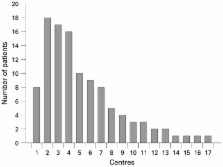
          The z score of the systemic ventricular end-diastolic dimension decreased by median 1.1 (p<0.001). Ejection fraction (EF) or fractional area of change increased by a mean (SD) of 11.5 (14.3)% (p<0.001) and New York Heart Association (NYHA) class improved by median 1.0 grade (p<0.001). Non-response to CRT (18.5%) was multivariably predicted by the presence of primary dilated cardiomyopathy (p = 0.002) and poor NYHA class (p = 0.003). Presence of a systemic left ventricle was the strongest multivariable predictor of improvement in EF/fractional area of change (p<0.001). Results were independent of the number of patients treated in each contributing centre.

          Conclusion:

          Heart failure associated with ventricular pacing is the largest indication for CRT in paediatric and congenital heart disease. CRT efficacy varies widely with the underlying anatomical and pathophysiological substrate.

          Related collections

          Most cited references19

          • Record: found
          • Abstract: found
          • Article: not found

          Adverse effect of ventricular pacing on heart failure and atrial fibrillation among patients with normal baseline QRS duration in a clinical trial of pacemaker therapy for sinus node dysfunction.

          Dual-chamber (DDDR) pacing preserves AV synchrony and may reduce heart failure (HF) and atrial fibrillation (AF) compared with ventricular (VVIR) pacing in sinus node dysfunction (SND). However, DDDR pacing often results in prolonged QRS durations (QRSd) as the result of right ventricular stimulation, and ventricular desynchronization may result. The effect of pacing-induced ventricular desynchronization in patients with normal baseline QRSd is unknown. Baseline QRSd was obtained from 12-lead ECGs before pacemaker implantation in MOST, a 2010-patient, 6-year, randomized trial of DDDR versus VVIR pacing in SND. Cumulative percent ventricular paced (Cum%VP) was determined from stored pacemaker data. Baseline QRSd 40%) and VVIR (HR 2.56 [95% CI, 1.48 to 4.43] for Cum%VP >80%). The risk of AF increased linearly with Cum%VP from 0% to 85% in both groups (DDDR, HR 1.36 [95% CI, 1.09, 1.69]; VVIR, HR 1.21 [95% CI 1.02, 1.43], for each 25% increase in Cum%VP). Model results were unaffected by adjustment for known baseline predictors of HF hospitalization and AF. Ventricular desynchronization imposed by ventricular pacing even when AV synchrony is preserved increases the risk of HF hospitalization and AF in SND with normal baseline QRSd.
            Bookmark
            • Record: found
            • Abstract: found
            • Article: not found

            Dual-chamber pacing or ventricular backup pacing in patients with an implantable defibrillator: the Dual Chamber and VVI Implantable Defibrillator (DAVID) Trial.

            Implantable cardioverter defibrillator (ICD) therapy with backup ventricular pacing increases survival in patients with life-threatening ventricular arrhythmias. Most currently implanted ICD devices provide dual-chamber pacing therapy. The most common comorbid cause for mortality in this population is congestive heart failure. To determine the efficacy of dual-chamber pacing compared with backup ventricular pacing in patients with standard indications for ICD implantation but without indications for antibradycardia pacing. The Dual Chamber and VVI Implantable Defibrillator (DAVID) Trial, a single-blind, parallel-group, randomized clinical trial. A total of 506 patients with indications for ICD therapy were enrolled between October 2000 and September 2002 at 37 US centers. All patients had a left ventricular ejection fraction (LVEF) of 40% or less, no indication for antibradycardia pacemaker therapy, and no persistent atrial arrhythmias. All patients had an ICD with dual-chamber, rate-responsive pacing capability implanted. Patients were randomly assigned to have the ICDs programmed to ventricular backup pacing at 40/min (VVI-40; n = 256) or dual-chamber rate-responsive pacing at 70/min (DDDR-70; n = 250). Maximal tolerated medical therapy for left ventricular dysfunction, including angiotensin-converting enzyme inhibitors and beta-blockers, was prescribed to all patients. Composite end point of time to death or first hospitalization for congestive heart failure. One-year survival free of the composite end point was 83.9% for patients treated with VVI-40 compared with 73.3% for patients treated with DDDR-70 (relative hazard, 1.61; 95% confidence interval [CI], 1.06-2.44). The components of the composite end point, mortality of 6.5% for VVI-40 vs 10.1% for DDDR-70 (relative hazard, 1.61; 95% CI, 0.84-3.09) and hospitalization for congestive heart failure of 13.3% for VVI-40 vs 22.6% for DDDR-70 (relative hazard, 1.54; 95% CI, 0.97-2.46), also trended in favor of VVI-40 programming. For patients with standard indications for ICD therapy, no indication for cardiac pacing, and an LVEF of 40% or less, dual-chamber pacing offers no clinical advantage over ventricular backup pacing and may be detrimental by increasing the combined end point of death or hospitalization for heart failure.
              Bookmark
              • Record: found
              • Abstract: found
              • Article: not found

              Detrimental ventricular remodeling in patients with congenital complete heart block and chronic right ventricular apical pacing.

              Although dual-chamber pacing improves cardiac function in patients with complete congenital atrioventricular block (CCAVB) by restoring physiological heart rate and atrioventricular synchronization, the long-term detrimental effect of asynchronous electromechanical activation induced by apical right ventricular pacing (RVP) has not been well clarified. Twenty-three CCAVB adults (24+/-3 years) with a DDD transvenous pacemaker underwent conventional echocardiography before implantation and, after at least 5 years of RVP, an exercise test and echocardiography coupled with tissue Doppler imaging and tissue tracking. They were compared with 30 matched healthy control subjects. After 10+/-3 years of RVP, CCAVB adults had significantly higher values versus controls in terms of intra-left ventricular (LV) asynchrony (respectively, 59+/-18 versus 19+/-9 ms, P<0.001), extent of LV myocardium displaying delayed longitudinal contraction (39+/-15% versus 10+/-7%, P<0.01), and septal-to-posterior wall-motion delay (84+/-26 versus 18+/-9 ms, P<0.01). The ratio of late-activated posterior to early-activated septal wall thickness was higher after long-term RVP than before (1.3+/-0.2 vs 1+/-0.1, P=0.05) and was higher than in controls (1+/-0.1, P<0.05). The percentage of patients with increased LV end-diastolic diameter was higher after long-term RVP than before implantation and was higher than in controls (57% versus 13%, P<0.05, and 57% versus 0%, P<0.01, respectively). CCAVB patients with long-term RVP had a lower cardiac output than controls (3.8+/-0.6 versus 4.9+/-0.8 L/min, P<0.05) and lower exercise performance (123+/-24 versus 185+/-39 W, P<0.001). Prolonged ventricular dyssynchrony induced by long-term endovenous RVP is associated with deleterious LV remodeling, LV dilatation, LV asymmetrical hypertrophy, and low exercise capacity. These new data highlight the importance of the ventricular activation sequence in all patients with chronic ventricular pacing.
                Bookmark

                Author and article information

                Journal
                Heart
                hrt
                Heart
                BMJ Publishing Group (BMA House, Tavistock Square, London, WC1H 9JR )
                1355-6037
                1468-201X
                2009
                15 July 2009
                22 March 2009
                : 95
                : 14
                : 1165-1171
                Affiliations
                [1 ]Department of Paediatric Cardiology, University of Leipzig, Heart Centre, Leipzig, Germany
                [2 ]Kardiocentrum and Cardiovascular Research Centre, University Hospital Motol, Prague, Czech Republic
                [3 ]German Heart Centre Berlin, Berlin, Germany
                [4 ]Bristol Royal Infirmary and University of Bristol, Bristol, UK
                [5 ]Hungarian Paediatric Heart Centre, Budapest, Hungary
                [6 ]Evelina Children’s Hospital, Guy’s and St Thomas’ Trust, London, UK
                [7 ]Département de Cardiologie Pédiatrique, Hôpital Necker, Paris, France
                [8 ]Paediatric Cardiology Unit, Rikshospitalet University Hospital, Oslo, Norway
                [9 ]Department of Paediatric Cardiology and Intensive Care, University Hospital, Göttingen, Germany
                [10 ]Department of Paediatric Cardiology, Leiden University Medical Centre, Leiden, The Netherlands
                [11 ]Division of Paediatric Cardiology, Department of Paediatrics, Helsinki University Central Hospital, Helsinki, Finland
                [12 ]Division of Paediatric Cardiology, University Children’s Hospital of Zurich, Zurich, Switzerland
                [13 ]Department of Paediatrics, Rigshospitalet, Copenhagen, Denmark
                [14 ]Beatrix Children’s Hospital, Division of Paediatric Cardiology, University Medical Centre Groningen, The Netherlands
                [15 ]Division of Paediatric Cardiology, AZ Maastricht, Maastricht, The Netherlands
                [16 ]Department of Paediatric Cardiology, Onassis Cardiac Surgery Centre, Athens, Greece
                [17 ]Servico de Cardiologia Pediatrica, Hospital de Santa Marta, Lisboa, Portugal
                Author notes
                Correspondence to: Professor J Janoušek, Department of Paediatric Cardiology, University of Leipzig, Heart Centre, Strümpellstrasse 39, 04289 Leipzig, Germany; jan.janousek@ 123456medizin.uni-leipzig.de
                Article
                ht160465
                10.1136/hrt.2008.160465
                2699215
                19307198
                66e2b060-6875-443c-9479-819a6b35ee4c
                © Janoušek et al 2009

                This is an open-access article distributed under the terms of the Creative Commons Attribution Non-commercial License, which permits unrestricted use, distribution, and reproduction in any medium, provided the original work is properly cited.

                History
                : 10 March 2009
                Categories
                Heart failure and cardiomyopathy
                Original articles
                1506

                Cardiovascular Medicine
                Cardiovascular Medicine

                Comments

                Comment on this article

                scite_

                Similar content318

                Cited by38

                Most referenced authors456