3
views
0
recommends
+1 Recommend
0 collections
    0
    shares
      • Record: found
      • Abstract: found
      • Article: found
      Is Open Access

      Hypertrophy of human embryonic stem cell–derived cardiomyocytes supported by positive feedback between Ca 2+ and diacylglycerol signals

      research-article

      Read this article at

      Bookmark
          There is no author summary for this article yet. Authors can add summaries to their articles on ScienceOpen to make them more accessible to a non-specialist audience.

          Abstract

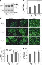
          Human embryonic stem cell–derived cardiomyocytes develop pronounced hypertrophy in response to angiotensin-2, endothelin-1, and a selected mix of three fatty acids. All three of these responses are accompanied by increases in both basal cytoplasmic Ca 2+ and diacylglycerol, quantified with the Ca 2+ sensor Fluo-4 and a FRET-based diacylglycerol sensor expressed in these cardiomyocytes. The heart glycoside, ouabain (30 nM), and a recently developed inhibitor of diacylglycerol lipases, DO34 (1 μM), cause similar hypertrophy responses, and both responses are accompanied by equivalent increases of basal Ca 2+ and diacylglycerol. These results together suggest that basal Ca 2+ and diacylglycerol form a positive feedback signaling loop that promotes execution of cardiac growth programs in these human myocytes. Given that basal Ca 2+ in myocytes depends strongly on the Na + gradient, we also tested whether nanomolar ouabain concentrations might stimulate Na +/K + pumps, as described by others, and thereby prevent hypertrophy. However, stimulatory effects of nanomolar ouabain (1.5 nM) were not verified on Na +/K + pump currents in stem cell–derived myocytes, nor did nanomolar ouabain block hypertrophy induced by endothelin-1. Thus, low-dose ouabain is not a “protective” intervention under the conditions of these experiments in this human myocyte model. To summarize, the major aim of this study has been to characterize the progression of hypertrophy in human embryonic stem cell–derived cardiac myocytes in dependence on diacylglycerol and Na + gradient changes, developing a case that positive feedback coupling between these mechanisms plays an important role in the initiation of hypertrophy programs.

          Related collections

          Most cited references75

          • Record: found
          • Abstract: found
          • Article: not found

          Autosis is a Na+,K+-ATPase-regulated form of cell death triggered by autophagy-inducing peptides, starvation, and hypoxia-ischemia.

          A long-standing controversy is whether autophagy is a bona fide cause of mammalian cell death. We used a cell-penetrating autophagy-inducing peptide, Tat-Beclin 1, derived from the autophagy protein Beclin 1, to investigate whether high levels of autophagy result in cell death by autophagy. Here we show that Tat-Beclin 1 induces dose-dependent death that is blocked by pharmacological or genetic inhibition of autophagy, but not of apoptosis or necroptosis. This death, termed "autosis," has unique morphological features, including increased autophagosomes/autolysosomes and nuclear convolution at early stages, and focal swelling of the perinuclear space at late stages. We also observed autotic death in cells during stress conditions, including in a subpopulation of nutrient-starved cells in vitro and in hippocampal neurons of neonatal rats subjected to cerebral hypoxia-ischemia in vivo. A chemical screen of ~5,000 known bioactive compounds revealed that cardiac glycosides, antagonists of Na(+),K(+)-ATPase, inhibit autotic cell death in vitro and in vivo. Furthermore, genetic knockdown of the Na(+),K(+)-ATPase α1 subunit blocks peptide and starvation-induced autosis in vitro. Thus, we have identified a unique form of autophagy-dependent cell death, a Food and Drug Administration-approved class of compounds that inhibit such death, and a crucial role for Na(+),K(+)-ATPase in its regulation. These findings have implications for understanding how cells die during certain stress conditions and how such cell death might be prevented.
            Bookmark
            • Record: found
            • Abstract: found
            • Article: not found

            Chemical and physiological characterization of fluo-4 Ca(2+)-indicator dyes.

            We have developed fluo-4, a new fluorescent dye for quantifying cellular Ca2+ concentrations in the 100 nM to 1 microM range. Fluo-4 is similar in structure and spectral properties to the widely used fluorescent Ca(2+)-indicator dye, fluo-3, but it has certain advantages over fluo-3. Due to its greater absorption near 488 nm, fluo-4 offers substantially brighter fluorescence emission when used with excitation by argon-ion laser or other sources in conjunction with the standard fluorescein filter set. In vitro, fluo-4 exhibited high fluorescence emission, a high rate of cell permeation, and a large dynamic range for reporting [Ca2+] around a Kd(Ca2+) of 345 nM. We have also developed several Ca(2+)-indicators related to fluo-4 having lower affinities for Ca2+ that are useful in cellular studies requiring quantification of higher [Ca2+]. In a variety of physiological studies of live cells, fluo-4 labeled cells more brightly than did fluo-3, when challenged with procedures designed to elevate calcium levels. Fluo-4 is well suited for photometric and imaging applications that make use of confocal laser scanning microscopy, flow cytometry, or spectrofluorometry, or in fluorometric high-throughput microplate screening assays. Because of its higher fluorescence emission intensity, fluo-4 can be used at lower intracellular concentrations, making its use a less invasive practice.
              Bookmark
              • Record: found
              • Abstract: found
              • Article: not found

              The C2 domain calcium-binding motif: structural and functional diversity.

              The C2 domain is a Ca(2+)-binding motif of approximately 130 residues in length originally identified in the Ca(2+)-dependent isoforms of protein kinase C. Single and multiple copies of C2 domains have been identified in a growing number of eukaryotic signalling proteins that interact with cellular membranes and mediate a broad array of critical intracellular processes, including membrane trafficking, the generation of lipid-second messengers, activation of GTPases, and the control of protein phosphorylation. As a group, C2 domains display the remarkable property of binding a variety of different ligands and substrates, including Ca2+, phospholipids, inositol polyphosphates, and intracellular proteins. Expanding this functional diversity is the fact that not all proteins containing C2 domains are regulated by Ca2+, suggesting that some C2 domains may play a purely structural role or may have lost the ability to bind Ca2+. The present review summarizes the information currently available regarding the structure and function of the C2 domain and provides a novel sequence alignment of 65 C2 domain primary structures. This alignment predicts that C2 domains form two distinct topological folds, illustrated by the recent crystal structures of C2 domains from synaptotagmin 1 and phosphoinositide-specific phospholipase C-delta 1, respectively. The alignment highlights residues that may be critical to the C2 domain fold or required for Ca2+ binding and regulation.
                Bookmark

                Author and article information

                Contributors
                Christine.deisl@utsouthwestern.edu
                214-645-6031 , Donald.hilgemann@utsouthwestern.edu
                Journal
                Pflugers Arch
                Pflugers Arch
                Pflugers Archiv
                Springer Berlin Heidelberg (Berlin/Heidelberg )
                0031-6768
                1432-2013
                28 June 2019
                28 June 2019
                2019
                : 471
                : 8
                : 1143-1157
                Affiliations
                ISNI 0000 0000 9482 7121, GRID grid.267313.2, Departments of Physiology and Internal Medicine, Charles and Jane Pak Center of Mineral Metabolism and Clinical Research, , University of Texas Southwestern Medical Center, ; 5323 Harry Hines Blvd, Dallas, TX 75235 USA
                Author information
                http://orcid.org/0000-0002-5288-5133
                Article
                2293
                10.1007/s00424-019-02293-0
                6614165
                31250095
                675beb39-f25a-423e-b10a-454c6f74d701
                © The Author(s) 2019

                Open Access This article is distributed under the terms of the Creative Commons Attribution 4.0 International License (http://creativecommons.org/licenses/by/4.0/), which permits unrestricted use, distribution, and reproduction in any medium, provided you give appropriate credit to the original author(s) and the source, provide a link to the Creative Commons license, and indicate if changes were made.

                History
                : 18 March 2019
                : 4 June 2019
                : 11 June 2019
                Funding
                Funded by: FundRef http://dx.doi.org/10.13039/100000002, National Institutes of Health;
                Award ID: HL119843
                Funded by: American Heart Associaton
                Award ID: Fellowship
                Award Recipient :
                Funded by: Charles and Jan Pak Foundation
                Award ID: N/A
                Categories
                Signaling and Cell Physiology
                Custom metadata
                © Springer-Verlag GmbH Germany, part of Springer Nature 2019

                Anatomy & Physiology
                diacylglycerol lipase,do34,cardiac hypertrophy,cardiac glycosides,ouabain,sodium potassium pump,patch clamp,angiotensin-2,endothelin-1

                Comments

                Comment on this article