19
views
0
recommends
+1 Recommend
0 collections
    0
    shares
      • Record: found
      • Abstract: found
      • Article: not found

      Mapping Sentinel Lymph Node Metastasis by Dual-probe Optical Imaging

      research-article

      Read this article at

      ScienceOpenPublisherPMC
      Bookmark
          There is no author summary for this article yet. Authors can add summaries to their articles on ScienceOpen to make them more accessible to a non-specialist audience.

          Abstract

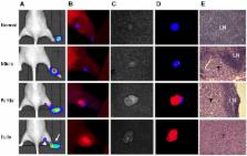
          Purpose: Sentinel lymph node biopsy (SLNB) has emerged as the preferred standard procedure in patients with breast cancer, melanoma and other types of cancer. Herein, we developed a method to intra-operatively map SLNs and differentiate tumor metastases within SLNs at the same time, with the aim to provide more accurate and real-time intraoperative guidance. Experimental Design: Hyaluronic acid (HA), a ligand of lymphatic vessel endothelial hyaluronan receptor (LYVE)-1, is employed as a SLN mapping agent after being conjugated with a near-infrared fluorophore (Cy5.5). Different sized HAs (5, 10 and 20K) were tested in normal mice and mice with localized inflammation to optimize LN retention time and signal to background ratio. Cetuximab, an antibody against epidermal growth factor receptor (EGFR), and trastuzumab, an antibody against human epidermal growth factor receptor 2 (HER2), were labeled with near-infrared fluorophore (IRDye800) for detecting metastatic tumors. LN metastasis model was developed by hock injection of firefly luciferase engineered human head neck squamous carcinoma cancer UM-SCC-22B cells or human ovarian cancer SKOV-3 cells. The metastases within LNs were confirmed by bioluminescence imaging (BLI). IRDye800-Antibodies were intravenously administered 24 h before local administration of Cy5.5-HA. Optical imaging was then performed to identify nodal metastases. Results: Binding of HA with LYVE-1 was confirmed by ELISA and fluorescence staining. HA with a size of 10K was chosen based on the favorable migration and retention profile. After sequential administration of IRDye800-antibodies intravenously and Cy5.5-HA locally to a mouse model with LN metastases and fluorescence optical imaging, partially metastasized LNs were successfully distinguished from un-metastasized LNs and fully tumor occupied LNs, based on the different signal patterns. Conclusions: Fluorophore conjugated HA is a potential lymphatic mapping agent for SLNB. Dual-tracer imaging with the combination of lymphatic mapping agents and tumor targeting agents can identify tumor metastases within SLNs, thus may provide accurate and real-time intra-operative guidance to spare the time spent waiting for a biopsy result.

          Related collections

          Most cited references33

          • Record: found
          • Abstract: found
          • Article: not found

          A comprehensive pathway map of epidermal growth factor receptor signaling

          The epidermal growth factor receptor (EGFR) signaling pathway is one of the most important pathways that regulate growth, survival, proliferation, and differentiation in mammalian cells. Reflecting this importance, it is one of the best-investigated signaling systems, both experimentally and computationally, and several computational models have been developed for dynamic analysis. A map of molecular interactions of the EGFR signaling system is a valuable resource for research in this area. In this paper, we present a comprehensive pathway map of EGFR signaling and other related pathways. The map reveals that the overall architecture of the pathway is a bow-tie (or hourglass) structure with several feedback loops. The map is created using CellDesigner software that enables us to graphically represent interactions using a well-defined and consistent graphical notation, and to store it in Systems Biology Markup Language (SBML).
            Bookmark
            • Record: found
            • Abstract: found
            • Article: not found

            The clinical use of indocyanine green as a near-infrared fluorescent contrast agent for image-guided oncologic surgery.

            Optical imaging using near-infrared (NIR) fluorescence provides new prospects for general and oncologic surgery. ICG is currently utilised in NIR fluorescence cancer-related surgery for three indications: sentinel lymph node (SLN) mapping, intraoperative identification of solid tumours, and angiography during reconstructive surgery. Therefore, understanding its advantages and limitations is of significant importance. Although non-targeted and non-conjugatable, ICG appears to be laying the foundation for more widespread use of NIR fluorescence-guided surgery. Copyright © 2011 Wiley-Liss, Inc.
              Bookmark
              • Record: found
              • Abstract: found
              • Article: not found

              Mechanisms of lymphatic metastasis.

              Malignant tumors release growth factors such as VEGF-C to induce lymphatic vessel expansion (lymphangiogenesis) in primary tumors and in draining sentinel LNs, thereby promoting LN metastasis. Surprising recent evidence suggests that lymphatic vessels do not merely represent passive channels for tumor spread, but that they may actively promote tumor cell recruitment to LNs, cancer stem cell survival, and immune modulation. New imaging approaches allow the sensitive visualization of the earliest LN metastases and the quantitative, noninvasive measurement of the function of tumor-draining lymphatic vessels, with potential applications in the development of biomarkers for prognosis and measurement of therapeutic response.
                Bookmark

                Author and article information

                Journal
                Theranostics
                Theranostics
                thno
                Theranostics
                Ivyspring International Publisher (Sydney )
                1838-7640
                2017
                1 January 2017
                : 7
                : 1
                : 153-163
                Affiliations
                [1 ]Jiangsu Key Laboratory of Molecular Imaging and Functional Imaging, Department of Radiology, Zhongda Hospital, Medical School of Southeast University, Nanjing 210009, China;
                [2 ]Laboratory of Molecular Imaging and Nanomedicine, National Institute of Biomedical Imaging and Bioengineering, National Institutes of Health (NIH), Bethesda, MD 20892, USA.
                Author notes
                ✉ Corresponding authors: Gao-Jun Teng, 87 Dingjiaqiao Road, Nanjing, Jiangsu, China, 210009, Email: gjteng@ 123456vip.sina.com Gang Niu, 35A Convent Drive Rm GD959, Bethesda, MD 20892, Email: niug@ 123456mail.nih.gov Xiaoyuan Chen, 35A Convent Drive Rm GD937, Bethesda, MD 20892, Email: shawn.chen@ 123456nih.gov .

                Competing Interests: The authors have declared that no competing interest exists.

                Article
                thnov07p0153
                10.7150/thno.17085
                5196893
                28042324
                67b68369-02e9-4728-adf6-f6f94a3be2cd
                © Ivyspring International Publisher. Reproduction is permitted for personal, noncommercial use, provided that the article is in whole, unmodified, and properly cited. See http://ivyspring.com/terms for terms and conditions.
                History
                : 2 August 2016
                : 4 October 2016
                Categories
                Research Paper

                Molecular medicine
                metastasis,sentinel lymph node,optical imaging,hyaluronic acid,egfr,her2.
                Molecular medicine
                metastasis, sentinel lymph node, optical imaging, hyaluronic acid, egfr, her2.

                Comments

                Comment on this article